. 2009 Mar 24;106(12):4707-12.
doi: 10.1073/pnas.0805963106. Epub 2009 Mar 2.

LOK is a major ERM kinase in resting lymphocytes and regulates cytoskeletal rearrangement through ERM phosphorylation

Affiliations

LOK is a major ERM kinase in resting lymphocytes and regulates cytoskeletal rearrangement through ERM phosphorylation

Natalya V Belkina et al. Proc Natl Acad Sci U S A. .

Abstract

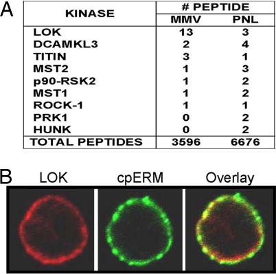
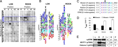
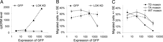
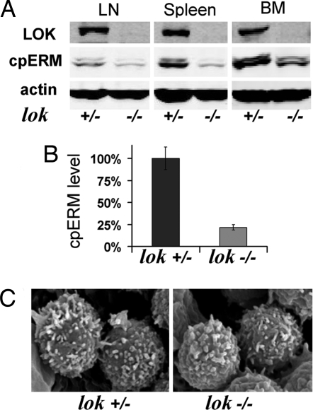
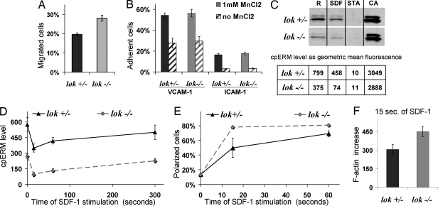
ERM (ezrin-radixin-moesin) proteins mediate linkage of actin cytoskeleton to plasma membrane in many cells. ERM activity is regulated in part by phosphorylation at a C-terminal threonine, but the identity of ERM kinases is unknown in lymphocytes and incompletely defined in other mammalian cells. Our studies show that lymphocyte-oriented kinase (LOK) is an ERM kinase in vitro and in vivo. Mass spectrometric analysis indicates LOK is abundant at the lymphocyte plasma membrane and immunofluorescence studies show LOK enrichment at the plasma membrane near ERM. In vitro peptide specificity analyses characterize LOK as a basophilic kinase whose optimal substrate sequence resembles the ERM site, including unusual preference for tyrosine at P-2. LOK's activity on moesin peptide and protein was comparable to reported ERM kinases ROCK and PKC but unlike them LOK displayed preferential specificity for moesin compared to traditional basophilic kinase substrates. Two genetic approaches demonstrate a role for LOK in ERM phosphorylation: cell transfection with LOK kinase domain augments ERM phosphorylation and lymphocytes from LOK knockout mice have >50% reduction in ERM phosphorylation. The findings on localization and specificity argue that LOK is a direct ERM kinase. The knockout mice have normal hematopoietic cell development but notably lymphocyte migration and polarization in response to chemokine are enhanced. These functional alterations fit the current understanding of the role of ERM phosphorylation in regulating cortical reorganization. Thus, these studies identify a new ERM kinase of importance in lymphocytes and confirm the role of ERM phosphorylation in regulating cell shape and motility.


Conflict of interest statement

The authors declare no conflict of interest.

Figures

Fig. 1.
Fig. 1.
LOK is abundant at the lymphocyte plasma membrane and colocalized with cpERM. (A) Tabulation of kinase peptides detected in mass spectrometric analysis of fractions from human lymphocytes. Fractions: MMV, membrane/microvillus; PNL, postnuclear lysate. (B) Colocalization of cpERM (green) and LOK (red) at human lymphocyte plasma membrane detected by immunofluorescence.
Fig. 2.
Fig. 2.
LOK has distinctive specificity for the ERM C-terminal phosphorylation site. (A) In vitro phosphorylation in solution of a degenerate peptide set (24) by LOK and ROCK. The intensity of each spot represents the amount of 32P incorporated into the corresponding degenerate peptide and therefore the kinase's preference for the particular residue at the indicated substrate position. Blue dotted frame and letters indicate basic amino acids. (B) The results from panel A are represented as a PSSM logo (25). Each stack of letters represents the kinase's preference for each possible residue at a single substrate position around the phosphorylation site (P0). Positions before P0 are represented as P minus (P–1, P–2) and after as P plus (P+1, P+2). The height of each letter is proportional to the absolute value of the log score of that preference and the positions of the letters in the stack are sorted from Bottom to Top in ascending value by the log score. Thus, strongly favored residues are at the Top and strongly disfavored residues are at the Bottom. Color indicates physicochemical properties of which the most relevant here are blue for basic, red for acidic, and black for hydrophobic. Arrow indicates P–2 position. (C) Sequences of the phosphorylation site in ERM from human, worm, and insect. Phosphorylation occurs on T and arrow indicates conserved Y at P–2. (D) Kinetic analysis of in vitro phosphorylation by LOK, ROCK, and PKC-θ of 2 peptides: WT moesin and mutant peptide where Y at P–2 is substituted with R (see also Fig. S3). (E) Phosphorylation of individual sites on 3 proteins by LOK (L), ROCK (R), and PKC-θ (P). Phosphorylated sites detected are: T558 on moesin C terminus (302–577), S10 on histone H3, and T850 on MYPT1 (654–880). Numbers to the Right of blots indicate level of fluorescent emissions from corresponding bands.
Fig. 3.
Fig. 3.
Evidence in Jurkat cells that LOK phosphorylates ERM and that ERM phosphorylation impedes migration. (A) Jurkat cells were transfected with LOK KD or GFP control and cpERM was detected by flow cytometry after 18 h. Cells from each preparation were separated into 4 categories by absolute GFP fluorescence i.e., nontransfected (0–10), low level expression (10–100), medium level expression (100–1000), and high level expression (>1000). (B) Same transfected cells as panel A analyzed for transmigration in response to SDF-1. (C) Jurkat cells were transfected with pseudophosphorylated (T558D), nonphosphorylated (T558A), or WT moesin GFP fusion protein and analyzed for transmigration in response to SDF-1.
Fig. 4.
Fig. 4.
Hematopoietic cells from LOK KO mice have decreased ERM phosphorylation without major change in lymphocyte microvilli. (A) Example of WB detection of cpERM, and actin in lysates from hematopoietic cells from different tissues, LN, lymph nodes; BM, bone marrow. (B) Comparison of cpERM level in spleen cells from 10 pairs of LOK KO mice and their heterozygote littermates. (C) SEM images of lymph node lymphocytes from a LOK KO mouse and its littermate.
Fig. 5.
Fig. 5.
LOK depletion results in alteration of migration and polarization, but not in adhesion. (A) Splenic lymphocytes from LOK KO mice and littermate controls were analyzed for transmigration to SDF-1. (B) Splenic lymphocytes from LOK KO mice and littermate controls were analyzed for adhesion to VCAM-1 or ICAM-1. Assays were performed in the presence or absence of 1 mM MnCl2, which promotes conversion of integrins into active conformation. (C) Splenic lymphocytes from LOK KO mice and littermate controls were analyzed for cpERM phosphorylation under 4 standard conditions: R, resting, no treatment; SDF-1, 1 min after 100 ng/mL SDF-1 to assess physiological dephosphorylation; STA, 5 min after high concentration (500 nM) staurosporine to assess maximal dephosphorylation; CA, 5 min after 50 μM phosphatase inhibitor calyculin A to maximize phosphorylation; cpERM was assessed both by WB (Upper subpanel) and flow cytometry (Lower subpanel). (D) cpERM levels were measured for splenic lymphocytes from LOK KO mice and littermate controls before and after various times of stimulation with SDF-1. (E) Cells as in panel D, but stained with phalloidin to detect F-actin and scored blind for polarization. (F) Cells as in panel E, but analyzed for increase in F-actin by flow cytometry after 15 s of stimulation.

References

    1. Charrin S, Alcover A. Role of ERM (ezrin-radixin-moesin) proteins in T lymphocyte polarization, immune synapse formation and in T cell receptor-mediated signaling. Front Biosci. 2006;11:1987–1997. - PubMed
    1. Niggli V, Rossy J. Ezrin/radixin/moesin: Versatile controllers of signaling molecules and of the cortical cytoskeleton. Int J Biochem Cell Biol. 2008;40:344–349. - PubMed
    1. Burkhardt JK, Carrizosa E, Shaffer MH. The actin cytoskeleton in T cell activation. Annu Rev Immunol. 2008;26:233–259. - PubMed
    1. Bretscher A, Edwards K, Fehon RG. ERM proteins and merlin: Integrators at the cell cortex. Nat Rev Mol Cell Biol. 2002;3:586–599. - PubMed
    1. Carreno S, et al. Moesin and its activating kinase Slik are required for cortical stability and microtubule organization in mitotic cells. J Cell Biol. 2008;180:739–746. - PMC - PubMed

Publication types

MeSH terms