

Cortical network functional connectivity in the descent to sleep
- PMID: 19255447
- PMCID: PMC2657465
- DOI: 10.1073/pnas.0900924106
Cortical network functional connectivity in the descent to sleep
Abstract
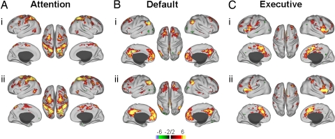
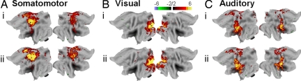
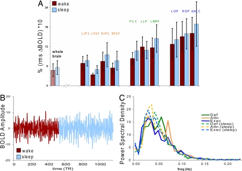
Descent into sleep is accompanied by disengagement of the conscious brain from the external world. It follows that this process should be associated with reduced neural activity in regions of the brain known to mediate interaction with the environment. We examined blood oxygen dependent (BOLD) signal functional connectivity using conventional seed-based analyses in 3 primary sensory and 3 association networks as normal young adults transitioned from wakefulness to light sleep while lying immobile in the bore of a magnetic resonance imaging scanner. Functional connectivity was maintained in each network throughout all examined states of arousal. Indeed, correlations within the dorsal attention network modestly but significantly increased during light sleep compared to wakefulness. Moreover, our data suggest that neuronally mediated BOLD signal variance generally increases in light sleep. These results do not support the view that ongoing BOLD fluctuations primarily reflect unconstrained cognition. Rather, accumulating evidence supports the hypothesis that spontaneous BOLD fluctuations reflect processes that maintain the integrity of functional systems in the brain.
Conflict of interest statement
The authors declare no conflict of interest.
Figures
References
-
- Rechtshaffen A, Kales A. A Manual of Standardized Terminology, Techniques, and Scoring System for Sleep Stages of Human Subjects. Univ. of California, Los Angeles: Brain Information Service/Brain Res Inst.; 1968.
-
- Iber C, Ancoli-Israel S, Chesson AL, Quan SF. The AASM Manual for the Scoring of Sleep and Associated Events. Westchester IL: American Academy of Sleep Medicine; 2007.
-
- Braun AR, et al. Regional cerebral blood flow throughout the sleep-wake cycle. Brain. 1997;120:1173–1197. - PubMed
-
- Maquet P. Functional neuroimaging of normal human sleep by positron emission tomography. J Sleep Res. 2000;9:207–231. - PubMed
-
- Kjaer TW, Law I, Wiltschiotz G, Paulson OB, Madsen PL. Regional cerebral blood flow during light sleep—A H(2)(15)O-PET study. J Sleep Res. 2002;11:201–207. - PubMed
Publication types
MeSH terms
Substances
Grants and funding
LinkOut - more resources
Full Text Sources
