. 2009 Jun;131(6):713-26.
doi: 10.1007/s00418-009-0577-1. Epub 2009 Mar 3.

Cellular organization of normal mouse liver: a histological, quantitative immunocytochemical, and fine structural analysis

Affiliations

Cellular organization of normal mouse liver: a histological, quantitative immunocytochemical, and fine structural analysis

Janie L Baratta et al. Histochem Cell Biol. 2009 Jun.

Abstract

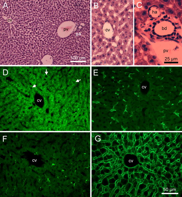
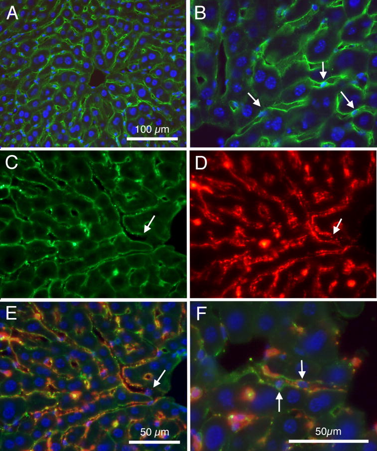
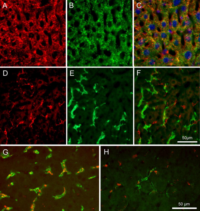
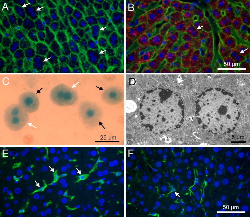
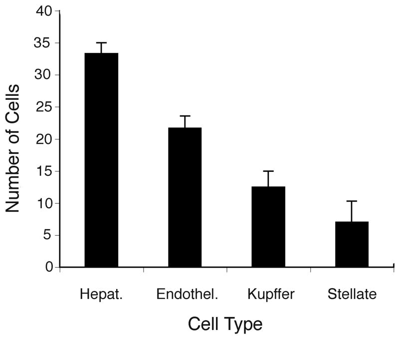
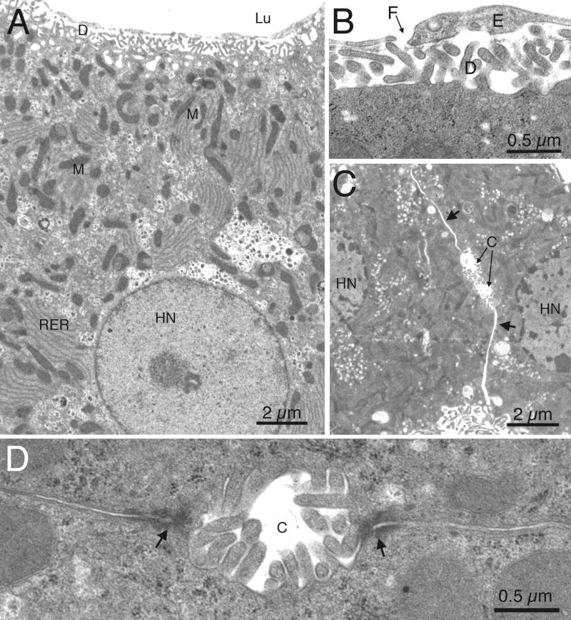
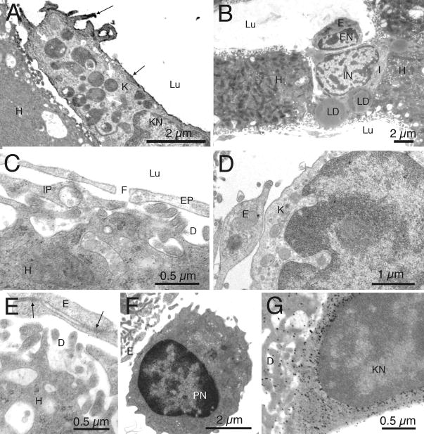
The cellular organization of normal mouse liver was studied using light and electron microscopy and quantitative immunocytochemical techniques. The general histological organization of the mouse liver is similar to livers of other mammalian species, with a lobular organization based on the distributions of portal areas and central venules. The parenchymal hepatocytes were detected with immunocytochemical techniques to recognize albumin or biotin containing cells. The macrophage Kupffer cells were identified with F4-80 immunocytochemistry, Ito stellate cells were identified with GFAP immunocytochemistry, and endothelial cells were labeled with the CD-34 antibody. Kupffer cells were labeled with intravascularly administered fluorescently labeled latex microspheres of both large (0.5 mum) and small (0.03 mum) diameters, while endothelial cells were labeled only with small diameter microspheres. Neither hepatocytes nor Ito stellate cells were labeled by intravascularly administered latex microspheres. The principal fine structural features of hepatocytes and non-parenchymal cells of mouse liver are similar to those reported for rat. Counts of immunocytochemically labeled cells with stained nuclei indicated that hepatocytes constituted approximately 52% of all labeled cells, Kupffer cells about 18%, Ito cells about 8%, and endothelial cells about 22% of all labeled cells. Approximately, 35% of the hepatocytes contained two nuclei; none of the Kupffer or Ito cells were double nucleated. The presence of canaliculi and a bile duct system appear similar to that reported for other species. The cellular organization of the mouse liver is quite similar to that of other mammalian species, confirming that the mouse presents a useful animal model for studies of liver structure and function.


Figures

Fig. 1
Fig. 1
Photomicrographs of 12 μm thick cryostat sections of mouse liver tissue. A: Bright field image of an H&E stained section showing normal liver architecture, and components of basic liver lobules, with portal area and central venule. B: Another H&E stained section at higher magnification. C: Bright field image showing profiles in a portal area, including branch of portal vein (pv), branch of hepatic artery (ha), and a small bile ductile (bd). D: Fluorescein optics showing Alexa 488 labeling of albumin immunoreactivity of hepatocytes; note the bright green packets of immunoreactivity in cytoplasm surrounding large round nuclei (white arrows) vacant of staining. E: Fluorescein optics showing Alexa 488 labeled F4-80 immunoreactivity of putative Kupffer cells. F: Alexa 488 labeled GFAP positive Ito stellate cells. G: Fluorescein labeled tomato lectin staining of cell borders including endothelial cells of sinusoidal capillaries. cv: central venule; pa: portal area; pv: portal venule. Calibration bar in ‘A’ = 100 μm; bar in ‘C’ = 25μm; bar in ‘G’ = 50 μm for ‘B’ and ‘D’ – ‘G’.
Fig. 2
Fig. 2
Fluorescence photomicrographs showing characteristics of endothelial cell labeling. A: Merged image showing Alexa 488 labeled CD-34 immunoreactivity (in green) and DAPI stained cell nuclei (in blue). B: Similar section as in ‘A’, but viewed at higher magnification. Note the many large round DAPI stained nuclei of hepatocytes. In addition, white arrows indicate several smaller and ovoid DAPI stained nuclei associated with the CD-34 positive endothelial cells. C: CD-34 immunoreactivity of endothelial cells of liver. D: Same section as in ‘C’ viewed under rhodamine optics, showing pattern of fluorescently labeled latex small (0.03 μm) microspheres. E: Same section as shown in ‘D’ and ‘E’, with merger of images viewed with fluorescein (green CD-34), rhodamine (red fluorescent latex microspheres) and ultraviolet (blue DAPI) optics. White arrow indicates the location of a putative endothelial cell nucleus, in ‘C’, ‘D’ and ‘E’. F: Merged image similar to ‘E’ but at higher magnification. Arrows indicate two putative endothelial cell nuclei. Calibration bar in A = 100μm. Bar in E = 50μm for C, D, and E. Bar in F = 50 μm for B and F.
Fig. 3
Fig. 3
Fluorescence photomicrographs showing cell specific labeling. A: Red Alexa 546 labeled antibody to albumin, demonstrating that virtually all cells containing round nuclei express albumin. B: Same section as ‘A’ viewed under fluorescein optics to show Alexa 488 labeled biotin immunocytochemistry. C: Merged images from ‘A’ and ‘B’, demonstrating that virtually all cells containing round nuclei appear co-labeled with albumin and biotin. D: Section processed with Alexa 546 labeled antibody to GFAP; note the labeled cells with stellate morphological features. E: Same section as shown in ‘D’, but viewed under fluorescein optics and showing F4-80 labeled cells. F: Merged images from ‘D’ and ‘E’, demonstrating the two immunocytochemical procedures label distinctly separate populations of cells. G: Section from an animal that received intravenous injections of rhodamine labeled fluorescent latex larage (0.5 μm) microspheres, and then processed for F4-80 immunocytochemistry. Note red microspheres associated with green F4-80 labeled cells. H: Section from same animal as ‘G’, but processed for GFAP immunocytochemistry. Note red microspheres appear separate from GFAP labeled cells. Calibration bar in F = 50μm for images A – F; calibration bar in H = 50 μm for G and H.
Fig. 4
Fig. 4
Evidence for double nucleated cells in liver tissue. A. Merged image shows green fluorescein labeled tomato lectin labeling of cell borders and blue DAPI staining of nuclei in a liver tissue section. White arrows indicate structures that appear to be cells with double nuclei. B: Merged image showing tissue labeled by a combination of tomato lectin binding (green) to reveal cell borders, and albumin immunocytochemistry (red) along with blue DAPI labeled nuclei. White arrows indicate several albumin positive cells within tomato lectin borders that display double nuclei. C: Trypan blue stained dissociated liver cells. Note three cells with single nuclei (black arrows) and two cells with double nuclei (white arrows). D: Electron micrograph of two nuclear profiles within a liver cell. E: Merged images, showing cells double labeled with F4-80 (green Alexa 488 label) and the blue DAPI labeled nuclei. F: Merged image showing cells double labeled with GFAP (green) and the blue DAPI labeled nuclei. White arrows in E and F indicate single nucleated cells. Calibration bar = 50 μm for ‘A’ and ‘B’, and for ‘E’ and ‘F’. Calibration bar in ‘C’ = 25 μm; bar in ‘D’ = 5 μm.
Fig. 5
Fig. 5
Histograms summarizing results of counts of immunocytochemically identified cells with DAPI stained nuclei. The Y axis shows numbers of cells detected in a region with an area of 48,000μm2, and thickness of 12 μm. Calculated numbers were adjusted using the method of Abercrombie (1946). Data are mean ± s.e.m.
Fig. 6
Fig. 6
Electron micrographs of liver tissue. A: Relatively low magnification electron micrograph showing a portion of an hepatocyte. A large round nucleus of the hepatocyte (HN) is seen in the lower part of the figure. Many profiles of rough endoplasmic reticulum (RER) can be seen throughout the cytoplasm, often intermixed with mitochondria (M). The space of Disse (D) can be seen at the basal surface, just below processes of endothelial cells that border the lumen (Lu). B: Relatively high magnification electron micrograph shows a fenestration (F) in the endothelial cell (E) lining, over the space of Disse (D). C: Electron micrograph demonstrating the inter-cellular space (arrows) and the presence of canaliculi between 2 adjacent hepatocytes. At this low magnification, intercellular occluding junctions are not visible. Nuclei of hepatocytes are indicated (HN). D: Higher magnification electron micrograph illustrating a canaliculus, and adjacent occluding junctions (arrows).
Fig. 7
Fig. 7
Electron micrographs of non-parenchymal cells. A: Electron micrograph of a section processed immunocytochemically for F4-80, showing cell surface immunoreactivity (black arrows) of a Kupffer cell (K); the nucleus of the Kupffer cell (KN) is visible. White arrows indicate lysomes within the Kupffer cell. The Kupffer cell is situated in the sinusoidal lumen (Lu), above an hepatocyte (H). B: Micrograph showing an Ito stellate cell (I) situated between two hepatocytes (H) and an endothelial cell (E). The Ito cell is characterized by cytoplasmic lipid droplets (LD). C: High magnification electron micrograph showing the space of Disse (D) situated under fenestrated (F) endothelial processes (EP) above an hepatocyte (H) and below the capillary lumen (Lu). Also seen is a putative Ito cell process (IP). D: Close membrane association, but without specialized membrane attachments, between a Kupffer cell (K) and an endothelial cell (E). E: Adhering junctions (arrows) between processes of 2 endothelial cells (E), overlying the space of Disse (D) and an hepatocyte (H). F: Pit cell with nucleus (PN) situated in a sinusoidal lumen, adjacent to an endothelial cell (E). G: Micrograph showing gold labeled particles associated with engulfed liposomes in a Kupffer cell (Kupffer cell nucleus (KN) is indicated).

Similar articles

Cited by

References

    1. Abercrombie M. Estimation of nuclear population from microtome sections. Anat Rec. 1946;94:239–247. - PubMed
    1. Aschoff L. Das Reticulo/endotheliale system. Ergebn Med Kinderheilk. 1924;26:1–118.
    1. Austyn JM, Gordon S. F4/80, a monoclonal antibody directed specifically against the mouse macrophage. Eur J Immunol. 1981;11:805–815. - PubMed
    1. Bartök I, Töth J, Remenar E, Viragh S. Fine structure of perisinusoidal cells in developing human and mouse liver. Acta Morphol Hung. 1983;31:337–352. - PubMed
    1. Bernuau D, Poliard A, Tournier I, Sala-Trepat J, Feldmann G. All hepatocytes are involved in the expression of the albumin gene in the normal adult rat: a demonstration by in situ hybridization and immunoperoxidase techniques. Cell Biol Int Rep. 1985;9:31–42. - PubMed

Publication types

MeSH terms

Substances