. 2009 Sep;104(5):511-22.
doi: 10.1007/s00395-009-0012-8. Epub 2009 Mar 3.

The cardiac sodium channel displays differential distribution in the conduction system and transmural heterogeneity in the murine ventricular myocardium

Affiliations

The cardiac sodium channel displays differential distribution in the conduction system and transmural heterogeneity in the murine ventricular myocardium

C A Remme et al. Basic Res Cardiol. 2009 Sep.

Abstract

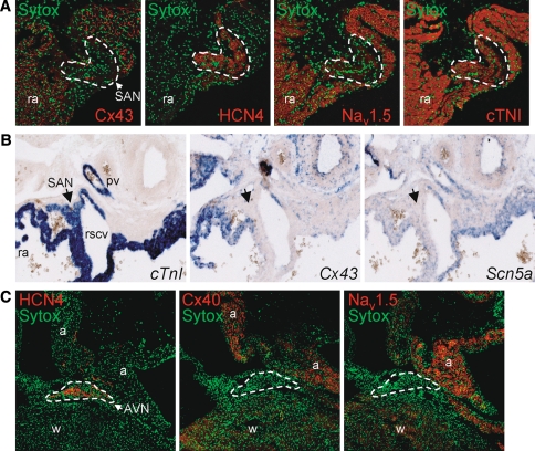
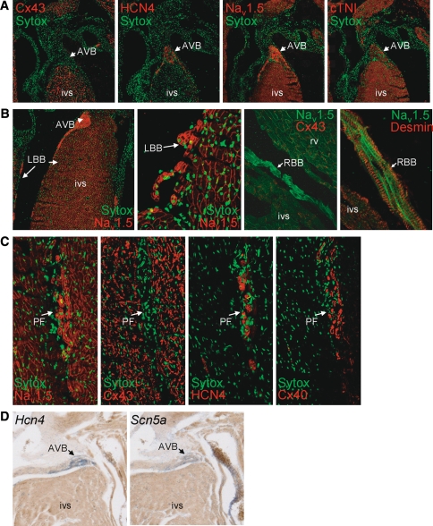
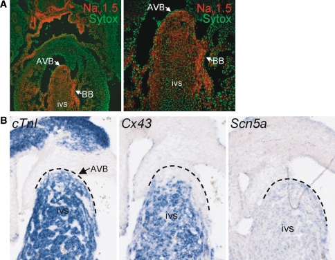
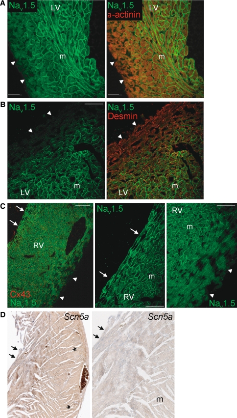
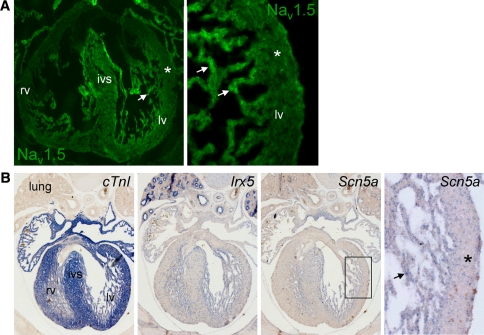
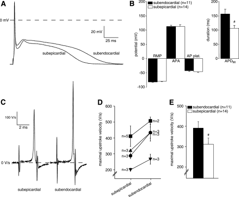
Cardiac sodium channels are responsible for conduction in the normal and diseased heart. We aimed to investigate regional and transmural distribution of sodium channel expression and function in the myocardium. Sodium channel Scn5a mRNA and Na(v)1.5 protein distribution was investigated in adult and embryonic mouse heart through immunohistochemistry and in situ hybridization. Functional sodium channel availability in subepicardial and subendocardial myocytes was assessed using patch-clamp technique. Adult and embryonic (ED14.5) mouse heart sections showed low expression of Na(v)1.5 in the HCN4-positive sinoatrial and atrioventricular nodes. In contrast, high expression levels of Na(v)1.5 were observed in the HCN4-positive and Cx43-negative AV or His bundle, bundle branches and Purkinje fibers. In both ventricles, a transmural gradient was observed, with a low Na(v)1.5 labeling intensity in the subepicardium as compared to the subendocardium. Similar Scn5a mRNA expression patterns were observed on in situ hybridization of embryonic and adult tissue. Maximal action potential upstroke velocity was significantly lower in subepicardial myocytes (mean +/- SEM 309 +/- 32 V/s; n = 14) compared to subendocardial myocytes (394 +/- 32 V/s; n = 11; P < 0.05), indicating decreased sodium channel availability in subepicardium compared to subendocardium. Scn5a and Na(v)1.5 show heterogeneous distribution patterns within the cardiac conduction system and across the ventricular wall. This differential distribution of the cardiac sodium channel may have profound consequences for conduction disease phenotypes and arrhythmogenesis in the setting of sodium channel disease.


Figures

Fig. 1
Fig. 1
a Immunohistochemical analysis of cryosections from adult murine heart showing a detailed view of the sinoatrial node (SAN; marked region). The SAN is clearly demarcated by the presence of HCN4 staining and the absence of Cx43 labeling (middle panel) and displays low levels of Nav1.5 staining. In contrast, abundant Nav1.5 staining is visible in the surrounding right atrial (ra) tissue. b In situ hybridization of embryonic heart (ED14.5) showing mRNA expression of Scn5a, CtnI and Cx43 (in blue). The CtnI-positive and Cx43-negative SAN (denoted by arrow) shows low Scn5a mRNA expression (pv pulmonary vein, rscv right superior caval vein). c Immunohistochemical analysis of cryosections from adult murine heart showing a detailed view of the atrioventricular node (AVN; marked region). The AVN shows intense staining for HCN4, but low expression of both Cx40 and Nav1.5, whereas clear Nav1.5 staining is visible in the atria (a) and working myocardium (w)
Fig. 2
Fig. 2
Distribution of Nav1.5 in the atrioventricular bundle (AVB) and His-Purkinje system in adult murine myocardium. a The AVB lacks Cx43 expression, is HCN4-positive, and shows a high Nav1.5 labeling intensity, continuing into (b) the left and right bundle braches, which show intense labeling of Nav1.5 and desmin, but absence of Cx43 (LBB left bundle branch, RBB right bundle branch, rv right ventricle, ivs intraventricular septum). c Serial sections of Purkinje fibers (PF) show high Nav1.5, HCN4, and Cx40 labeling intensity, but no Cx43. d In situ hybridization of adult heart shows similar high expression levels of Hcn4 and Scn5a mRNA in AVB (blue signals) in comparison with the myocardium of the intraventricular septum (ivs)
Fig. 3
Fig. 3
Distribution of Nav1.5 and Scn5a in the conduction system in embryonic hearts (ED14.5). a High expression levels of Nav1.5 protein in the AV bundle (AVB) and bundle branches (BB) in comparison with the ventricular myocardium of the intraventricular septum (ivs); the right panel represents an enlarged section of the left panel. b In situ hybridization shows prominent Scn5a mRNA expression (in blue) but absence of Cx43 mRNA in the AVB (dashed line indicates region of AVB)
Fig. 4
Fig. 4
Transmural distribution of Nav1.5 in the ventricular myocardium. Low Nav1.5 labeling intensity in ventricular epicardium (arrowheads) compared to the midmyocardial layer (m) in (a) the apex and (b) left ventricular free wall of the adult mouse heart. For comparison, α-actinin and desmin show a homogeneous transmural labeling. cLeft panel shows low Nav1.5 expression in subepicardium (arrowheads) and high Nav1.5 expression in subendocardium (arrows) of the right ventricle. For comparison, Cx43 shows a homogeneous transmural distribution. Also shown are details of subendocardial surface (middle panel) and subepicardial region (right panel) of the right ventricle. d In situ hybridization of adult ventricular myocardium shows clear Scn5a staining (in blue) in the subendocardium (arrows) and low Scn5a expression in the subepicardium (asterisks). The right panel depicts a magnified section of the left panel indicating higher Scn5a expression in the subendocardium (arrows) compared to midmural (m)
Fig. 5
Fig. 5
a Overview (left panel) and detail (right panel) of embryonic mouse heart (ED14.5) indicating low Nav1.5 labeling intensity in ventricular subepicardium (asterisks) and abundant Nav1.5 in the trabeculated subendocardium. b In situ hybridization of ventricular myocardium in embryonic heart (ED14.5; overview and magnified boxed section in the right panel) displaying a transmural gradient in Scn5a mRNA expression with lower expression in the subepicardium (asterisks) compared to the trabeculated subendocardium (arrows). For comparison, the homeodomain transcription factor Irx5 shows a similar transmural distribution pattern (lv left ventricle, rv right ventricle, ivs intraventricular septum)
Fig. 6
Fig. 6
a Representative examples of action potentials of a subepicardial and subendocardial myocyte from one heart, measured at a stimulation frequency of 2 Hz. b Averaged action potential characteristics (RMP resting membrane potential, APA action potential amplitude, AP plat. action potential plateau phase 20 ms after the upstroke, APD90 action potential duration at 90% repolarization, #P < 0.05 versus subendocardial). c Representative examples of the first time-derivative of the action potential (dV/dtmax), reflecting the maximal action potential upstroke velocity, measured from a subepicardial and subendocardial myocyte from one heart at a stimulation frequency of 2 Hz. d Averaged subepicardial and subendocardial action potential upstroke velocities of each heart measured (n = 4 hearts); numbers of mycoytes measured from each heart are indicated. e Average values for action potential upstroke velocities from all subepicardial and subendocardial myocytes (#P < 0.05 versus subendocardial)

References

    1. {'text': '', 'ref_index': 1, 'ids': [{'type': 'DOI', 'value': '10.1007/s003950170002', 'is_inner': False, 'url': 'https://doi.org/10.1007/s003950170002'}, {'type': 'PubMed', 'value': '11770069', 'is_inner': True, 'url': 'https://pubmed.ncbi.nlm.nih.gov/11770069/'}]}
    2. Antzelevitch C, Fish J (2001) Electrical heterogeneity within the ventricular wall. Basic Res Cardiol 96:517–527 - PubMed
    1. {'text': '', 'ref_index': 1, 'ids': [{'type': 'DOI', 'value': '10.1016/S0022-0736(99)90074-2', 'is_inner': False, 'url': 'https://doi.org/10.1016/s0022-0736(99)90074-2'}, {'type': 'PubMed', 'value': '10688320', 'is_inner': True, 'url': 'https://pubmed.ncbi.nlm.nih.gov/10688320/'}]}
    2. Antzelevitch C, Yan GX, Shimizu W (1999) Transmural dispersion of repolarization and arrhythmogenicity: the Brugada syndrome versus the long QT syndrome. J Electrocardiol 32(Suppl):158–165 - PubMed
    1. {'text': '', 'ref_index': 1, 'ids': [{'type': 'DOI', 'value': '10.1111/j.1469-7793.2001.0439c.xd', 'is_inner': False, 'url': 'https://doi.org/10.1111/j.1469-7793.2001.0439c.xd'}, {'type': 'PMC', 'value': 'PMC2278873', 'is_inner': False, 'url': 'https://pmc.ncbi.nlm.nih.gov/articles/PMC2278873/'}, {'type': 'PubMed', 'value': '11600679', 'is_inner': True, 'url': 'https://pubmed.ncbi.nlm.nih.gov/11600679/'}]}
    2. Ashamalla SM, Navarro D, Ward CA (2001) Gradient of sodium current across the left ventricular wall of adult rat hearts. J Physiol 536:439–443 - PMC - PubMed
    1. {'text': '', 'ref_index': 1, 'ids': [{'type': 'DOI', 'value': '10.1152/ajpheart.91495.2007', 'is_inner': False, 'url': 'https://doi.org/10.1152/ajpheart.91495.2007'}, {'type': 'PMC', 'value': 'PMC2494753', 'is_inner': False, 'url': 'https://pmc.ncbi.nlm.nih.gov/articles/PMC2494753/'}, {'type': 'PubMed', 'value': '18456723', 'is_inner': True, 'url': 'https://pubmed.ncbi.nlm.nih.gov/18456723/'}]}
    2. Bébarová M, O’Hara T, Geelen JL, Jongbloed RJ, Timmermans C, Arens YH, Rodriguez LM, Rudy Y, Volders PG (2008) Subepicardial phase 0 block and discontinuous transmural conduction underlie right precordial ST-segment elevation by a SCN5A loss-of-function mutation. Am J Physiol Heart Circ Physiol 295:H48–H58 - PMC - PubMed
    1. {'text': '', 'ref_index': 1, 'ids': [{'type': 'PMC', 'value': 'PMC198523', 'is_inner': False, 'url': 'https://pmc.ncbi.nlm.nih.gov/articles/PMC198523/'}, {'type': 'PubMed', 'value': '14523039', 'is_inner': True, 'url': 'https://pubmed.ncbi.nlm.nih.gov/14523039/'}]}
    2. Benson DW, Wang DW, Dyment M, Knilans TK, Fish FA, Strieper MJ, Rhodes TH, George AL Jr (2003) Congenital sick sinus syndrome caused by recessive mutations in the cardiac sodium channel gene (SCN5A). J Clin Invest 112:1019–1028 - PMC - PubMed

Publication types

MeSH terms