. 2009 Jun;5(2):139-49.
doi: 10.1007/s11302-009-9135-5. Epub 2009 Mar 3.

Activation of the P2X7 ion channel by soluble and covalently bound ligands

Affiliations

Activation of the P2X7 ion channel by soluble and covalently bound ligands

Nicole Schwarz et al. Purinergic Signal. 2009 Jun.

Abstract

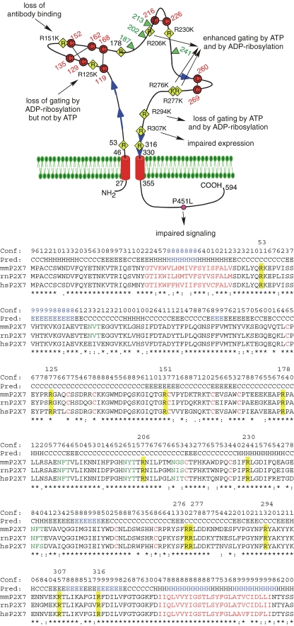
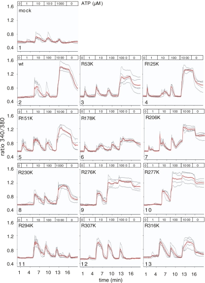
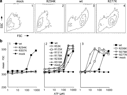
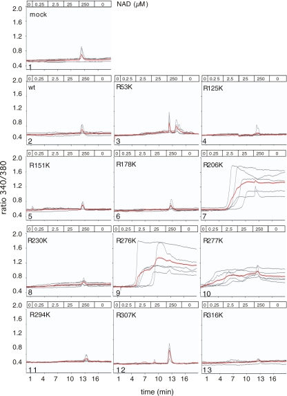
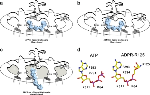
The homotrimeric P2X7 purinergic receptor has sparked interest because of its capacity to sense adenosine triphosphate (ATP) and nicotinamide adenine dinucleotide (NAD) released from cells and to induce calcium signaling and cell death. Here, we examine the response of arginine mutants of P2X7 to soluble and covalently bound ligands. High concentrations of ecto-ATP gate P2X7 by acting as a soluble ligand and low concentrations of ecto-NAD gate P2X7 following ADP-ribosylation at R125 catalyzed by toxin-related ecto-ADP-ribosyltransferase ART2.2. R125 lies on a prominent cysteine-rich finger at the interface of adjacent receptor subunits, and ADP-ribosylation at this site likely places the common adenine nucleotide moiety into the ligand-binding pocket of P2X7.


Figures

Fig. 1
Fig. 1
Schematic diagram of the functional consequences following substitution of the conserved arginine residues in the ectodomain of mouse P2X7. The connectivity of cysteine residues (in red) corresponds to that proposed for P2X1 and P2X2 [6]. The conserved arginine (R) residues in the ectodomain are indicated by yellow diamonds, the natural allelic polymorphism in the cytosolic domain [70] that distinguishes C57BL/6 mice from wild-type mice is indicated by a pink circle. Potential glycosylation sites are indicated by green triangles and predicted β-strands by blue arrows. The sequences in the alignment of mouse, rat, and human P2X7 are truncated five residues downstream of Tm2. Secondary structures predicted with PSIPRED [49] are indicated above the alignment (H helix; E extended β-strand; C coil, unstructured). Structure units with a confidence >8 are highlighted in blue. Identical, strongly and weakly conserved amino acid residues are indicated by asterisk and colon. Predicted transmembrane domains and conserved cysteine residues are in red, potential N-linked glycosylation sites are in green. The 11 conserved arginine residues in the ectodomain are highlighted in yellow and their positions in residue number (for mouse P2X7) are indicated above the alignment
Fig. 2
Fig. 2
Potency of ATP to induce calcium flux in HEK cells transiently transfected with P2X7 variants. HEK cells were co-transfected with expression constructs for mRFP and wild-type or mutant P2X7 purinoceptors. Twenty hours post-transfection, cells were loaded with the calcium-sensitive fluorochrome Fura-2 before live cell imaging by fluorescence microscopy. Images were captured every 5 s. At the indicated times, the perfusion buffer (37°C) was changed to subject cells to increasing doses of ATP. Ratio images (340/380 nm) were constructed pixel-by-pixel and single cell tracings were captured using the Openlab software. Gray lines show single cell tracings, red lines the calculated mean
Fig. 3
Fig. 3
Potency of ATP to induce changes in forward and side scatter of HEK cells transiently transfected with P2X7 variants. HEK cells were transfected with expression constructs for wild-type or mutant P2X7 receptors. Twenty hours post-transfection, cells were harvested by mild trypsinization and cells were incubated for 60 min in the absence or presence of the indicated concentrations of ATP before FACS analyses. a Contour plots illustrating the ATP-induced changes in cell size (forward scatter, FSC) and cell granularity (side scatter, SSC). b Dose–response curves illustrating the mean FSC of HEK cells treated as in (a) as a function of the concentration of ATP
Fig. 4
Fig. 4
Potency of ADP-ribosylation to induce calcium flux in HEK cells transiently transfected with P2X7 variants. HEK cells were co-transfected with expression constructs for mRFP, ART2.2, and wild-type or mutant P2X7 purinoceptors. Twenty hours post-transfection, cells were loaded with the calcium-sensitive fluorochrome Fura-2 before live cell imaging by fluorescence microscopy as in Fig. 2. At the indicated times, the perfusion buffer (37°C) was changed to subject cells to increasing doses of NAD. Gray lines show single cell tracings, red lines the calculated mean
Fig. 5
Fig. 5
Model for the activation of P2X7 by ADP-ribosylation at R125. a Schematic diagram of the trimeric P2X7 receptor complex in open conformation following binding of ATP at the interface of two adjacent subunits. b ADP-ribosylation at R125 places the attached ADP-ribose in the ligand-binding site, inducing the open conformation. c ADP-ribosylation at residue R133 places the attached ADP-ribose out of reach of the binding site. d Schematic diagrams of the soluble ligand ATP and of ADP-ribose in covalent linkage to R125. Residues K64 and K311 may interact with the negatively charged phosphate groups, residues F293 and R294 with the adenine–ribose moiety [6, 76]
Fig. 6
Fig. 6
Summary of mutagenesis data for P2X receptors in the region of the cysteine-rich finger. The arginine residues that serve as targets for ADP-ribosylation in mouse P2X7 [23], rat, and human P2X7 (our own unpublished observations) are highlighted in cyan. Conserved cysteine residues are highlighted in yellow. Amino acid residues that, when mutated, result in a gain-of-function are shown in bold and are highlighted in green [59, 60]. Residues that alter the sensitivity of the receptor to zinc, copper, and/or magnesium are shown in bold red [58, 59, 61, 77]. The positions of the cysteines (numbering for mouse P2X7) are indicated on top, the proposed connectivity [6] is indicated below. mm = Mus musculus, hs = Homo sapiens, rn = Rattus norvegicus, sm = Schistosoma mansoni, dd = Dictyostelium discoideum

Similar articles

Cited by

References

    1. {'text': '', 'ref_index': 1, 'ids': [{'type': 'DOI', 'value': '10.1046/j.1432-1327.2000.01187.x', 'is_inner': False, 'url': 'https://doi.org/10.1046/j.1432-1327.2000.01187.x'}, {'type': 'PubMed', 'value': '10712584', 'is_inner': True, 'url': 'https://pubmed.ncbi.nlm.nih.gov/10712584/'}]}
    2. Ziegler M (2000) New functions of a long-known molecule. Emerging roles of NAD in cellular signaling. Eur J Biochem 267:1550–1564 - PubMed
    1. {'text': '', 'ref_index': 1, 'ids': [{'type': 'DOI', 'value': '10.1189/jlb.0802418', 'is_inner': False, 'url': 'https://doi.org/10.1189/jlb.0802418'}, {'type': 'PubMed', 'value': '12629147', 'is_inner': True, 'url': 'https://pubmed.ncbi.nlm.nih.gov/12629147/'}]}
    2. la Sala A, Ferrari D, Di Virgilio F, Idzko M, Norgauer J, Girolomoni G (2003) Alerting and tuning the immune response by extracellular nucleotides. J Leukoc Biol 73:339–343 - PubMed
    1. {'text': '', 'ref_index': 1, 'ids': [{'type': 'DOI', 'value': '10.1038/nature04886', 'is_inner': False, 'url': 'https://doi.org/10.1038/nature04886'}, {'type': 'PubMed', 'value': '16885977', 'is_inner': True, 'url': 'https://pubmed.ncbi.nlm.nih.gov/16885977/'}]}
    2. Khakh BS, North RA (2006) P2X receptors as cell-surface ATP sensors in health and disease. Nature 442:527–532 - PubMed
    1. {'text': '', 'ref_index': 1, 'ids': [{'type': 'DOI', 'value': '10.1182/blood.V97.3.587', 'is_inner': False, 'url': 'https://doi.org/10.1182/blood.v97.3.587'}, {'type': 'PubMed', 'value': '11157473', 'is_inner': True, 'url': 'https://pubmed.ncbi.nlm.nih.gov/11157473/'}]}
    2. Di Virgilio F, Chiozzi P, Ferrari D, Falzoni S, Sanz JM, Morelli A, Torboli M, Bolognesi G, Baricordi OR (2001) Nucleotide receptors: an emerging family of regulatory molecules in blood cells. Blood 97:587–600 - PubMed
    1. {'text': '', 'ref_index': 1, 'ids': [{'type': 'PubMed', 'value': '12270951', 'is_inner': True, 'url': 'https://pubmed.ncbi.nlm.nih.gov/12270951/'}]}
    2. North RA (2002) Molecular physiology of P2X receptors. Physiol Rev 82:1013–1067 - PubMed

LinkOut - more resources