Review
. 2008:24:71-87.
doi: 10.1016/j.yadr.2008.09.004.

Toll-like receptors in skin

Affiliations
Review

Toll-like receptors in skin

Lloyd S Miller. Adv Dermatol. 2008.

Abstract

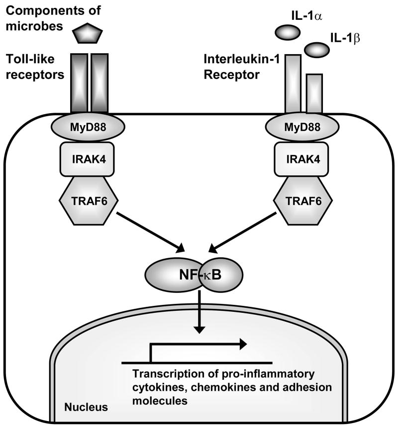
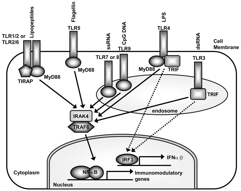
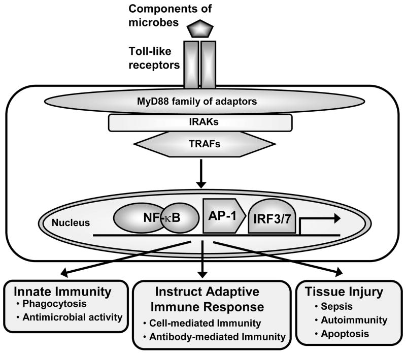
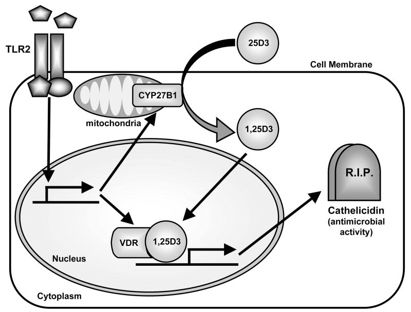
TLRs have emerged as a major class of PRRs that are involved in detecting invading pathogens in the skin and initiating cutaneous immune responses. TLRs are expressed on many different cell types in the skin, including keratinocytes and Langerhans cells in the epidermis. Each TLR can recognize a different microbial component and there are differences among the TLR signaling pathways, which lead to distinct immune responses against a given pathogen. Certain TLRs have been implicated in the pathogenesis of skin diseases, such as atopic dermatitis, psoriasis, and acne vulgaris. In addition, TLRs have been shown to be important in cutaneous host defense mechanisms against common bacterial, fungal, and viral pathogens in the skin, such as S aureus, C albicans, and HSV. Since the discovery that topical TLR agonists promote antiviral and antitumor immune responses, there has been considerable interest in the development of TLR-based therapies for skin diseases, skin cancer, and infections. Future research involving TLRs in skin will hopefully provide new insights into host defense against skin pathogens and novel therapeutic targets aimed at treating skin disease and skin cancer.


Figures

Figure 1
Figure 1. TLRs and the interleukin-1 receptor share a similar signaling cascade to initiate immune responses
TLRs and the interleukin 1 receptor (IL-1R) share a similar signaling cascade, which involve activation of the adapter molecule MyD88. MyD88 forms an initial signaling complex with IRAK4 and TRAF6. Formation of this complex results in activation of a signaling cascade that eventually leads to activation of NF-κB (and other pathways) to promote transcription of pro-inflammatory cytokines, chemokines and co-stimulatory and adhesion molecules involved in innate and adaptive immune responses.
Figure 2
Figure 2. Pathogen-associated molecular patterns (PAMPs) recognized by TLRs, the cellular location of TLRs and the different MyD88 adapters used by TLRs that promote distinct immune responses
Each TLR recognizes a different microbial component. TLR2 forms a heterodimer with TLR1 or TLR6 to recognize tri- and di-acyl lipopeptides, respectively. TLR4 recognizes LPS and TLR5 recognizes flagellin. These TLRs are located on the cell membrane and become internalized into phagosomes after interaction with their ligands. In contrast, TLR3 recognizes viral dsRNA, TLR7 and TLR8 recognize viral ssRNA, and TLR9 recognizes hypomethylated DNA (CpG motifs) of both bacteria and viruses and are located in intracellular membranes of endosomes and lysosomes. TLRs utilize MyD88 and TRIF adapters to initiate signaling. All TLRs except TLR3 can signal via MyD88. TLR2 and TLR4 also require the presence of TIRAP. MyD88 initiates a signaling cascade that eventually results in activation of NF-κB (and other pathways) to promote transcription of immunomodulatory genes. In contrast, TLR3 and TLR4 can also signal via TRIF in a MyD88-independent pathway. The TRIF pathway is critical in activating IRF3 (and IRF7), which promotes production of type I interferon (i.e. IFNα and IFNβ) and anti-viral immune responses.
Figure 3
Figure 3. Immune responses generated by activation of TLRs
TLRs recognize various microbial components and transduce signals via the family of MyD88 adapter molecules. MyD88 adapters recruit interleukin-1 receptor-associated kinases (IRAKs) and tumor necrosis factor receptor-associated factors (TRAFs) to form the initial signaling complexes that lead to activation of downstream signaling pathways, including activation of transcription factors such as NF-κB, AP-1 (activator protein-1), and IRF3/7 (interferon regulatory factors 3 and 7). These signaling pathways are responsible for distinct gene programs involved in different innate and acquired immune responses. TLRs have also been implicated in tissue injury in conditions such as sepsis, autoimmunity, and apoptosis.
Figure 4
Figure 4. TLR2 activates a vitamin D-dependent antimicrobial pathway
Activation of TLR2 on human monocytes/macrophages or keratinocytes from healing wounds (or keratinocytes stimulated with TGF-β) in increased expression of the vitamin D-1-hydroxylase CYP27B1 and the vitamin D receptor (VDR). CYP27B1 converts the inactive form of vitamin D (25D3) to its active form (1,25D3). 1,25D3 binds to and activates the VDR, which induces production of the antimicrobial peptide cathelicidin. Since cathelicidin has microbicidal activity against a variety of pathogenic microorganisms, TLR2 induction of a vitamin D-dependent antimicrobial pathway may be an important mechanism for cutaneous host defense.

Similar articles

Cited by

References

    1. Kang SS, Kauls LS, Gaspari AA. Toll-like receptors: applications to dermatologic disease. J Am Acad Dermatol. 2006;54(6):951–983. - PubMed
    1. McInturff JE, Modlin RL, Kim J. The role of toll-like receptors in the pathogenesis and treatment of dermatological disease. J Invest Dermatol. 2005;125(1):1–8. - PubMed
    1. Miller LS, Modlin RL. Toll-like receptors in the skin. Semin Immunopathol. 2007;29(1):15–26. - PubMed
    1. Clark R, Kupper T. Old meets new: the interaction between innate and adaptive immunity. J Invest Dermatol. 2005;125(4):629–637. - PubMed
    1. Kupper TS, Fuhlbrigge RC. Immune surveillance in the skin: mechanisms and clinical consequences. Nat Rev Immunol. 2004;4(3):211–222. - PMC - PubMed

Substances