. 2009 Apr;22(4):699-707.
doi: 10.1021/tx800464q.

Serum metabolomics reveals irreversible inhibition of fatty acid beta-oxidation through the suppression of PPARalpha activation as a contributing mechanism of acetaminophen-induced hepatotoxicity

Affiliations

Serum metabolomics reveals irreversible inhibition of fatty acid beta-oxidation through the suppression of PPARalpha activation as a contributing mechanism of acetaminophen-induced hepatotoxicity

Chi Chen et al. Chem Res Toxicol. 2009 Apr.

Abstract

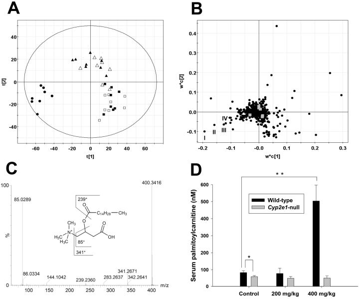
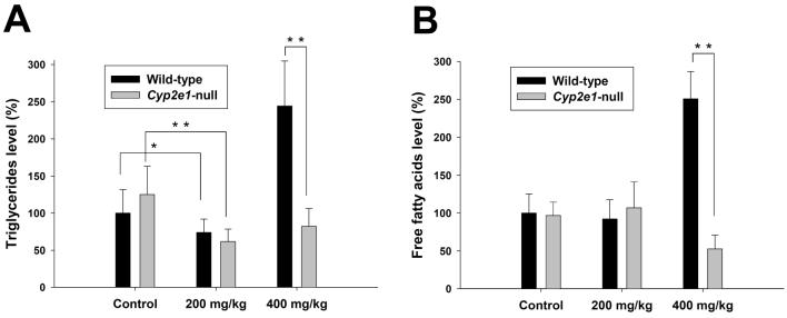
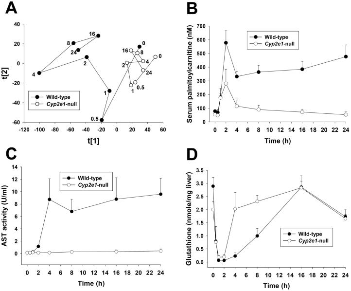
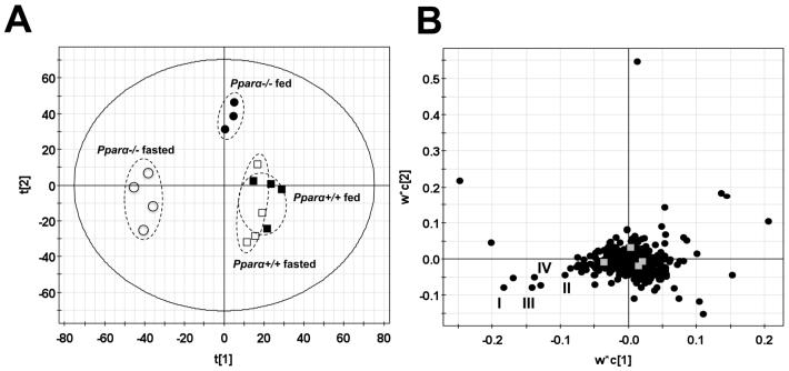
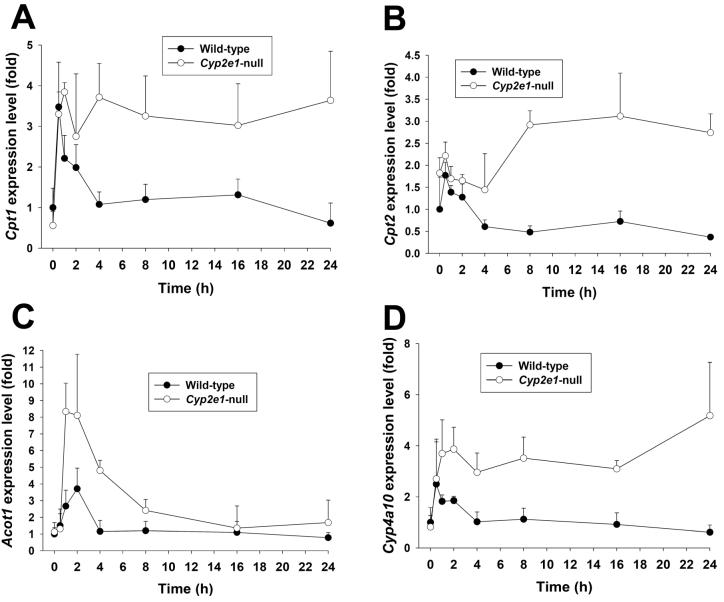
Metabolic bioactivation, glutathione depletion, and covalent binding are the early hallmark events after acetaminophen (APAP) overdose. However, the subsequent metabolic consequences contributing to APAP-induced hepatic necrosis and apoptosis have not been fully elucidated. In this study, serum metabolomes of control and APAP-treated wild-type and Cyp2e1-null mice were examined by liquid chromatography-mass spectrometry (LC-MS) and multivariate data analysis. A dose-response study showed that the accumulation of long-chain acylcarnitines in serum contributes to the separation of wild-type mice undergoing APAP-induced hepatotoxicity from other mouse groups in a multivariate model. This observation, in conjunction with the increase of triglycerides and free fatty acids in the serum of APAP-treated wild-type mice, suggested that APAP treatment can disrupt fatty acid beta-oxidation. A time-course study further indicated that both wild-type and Cyp2e1-null mice had their serum acylcarnitine levels markedly elevated within the early hours of APAP treatment. While remaining high in wild-type mice, serum acylcarnitine levels gradually returned to normal in Cyp2e1-null mice at the end of the 24 h treatment. Distinct from serum aminotransferase activity and hepatic glutathione levels, the pattern of serum acylcarnitine accumulation suggested that acylcarnitines can function as complementary biomarkers for monitoring the APAP-induced hepatotoxicity. An essential role for peroxisome proliferator-activated receptor alpha (PPARalpha) in the regulation of serum acylcarnitine levels was established by comparing the metabolomic responses of wild-type and Ppara-null mice to a fasting challenge. The upregulation of PPARalpha activity following APAP treatment was transient in wild-type mice but was much more prolonged in Cyp2e1-null mice. Overall, serum metabolomics of APAP-induced hepatotoxicity revealed that the CYP2E1-mediated metabolic activation and oxidative stress following APAP treatment can cause irreversible inhibition of fatty acid oxidation, potentially through suppression of PPARalpha-regulated pathways.


Figures

Figure 1
Figure 1
Identification of acylcarnitines as serum biomarkers of APAP toxicity through metabolomic analysis of 24-h serum samples from wild-type and Cyp2e1-null mice treated with 200 and 400 mg/kg APAP. A, Scores scatter plot of an PLS-DA model on the APAP-elicited dose-dependent influence on the serum metabolome. Details of data processing and model construction were described in the Experimental procedures. A two-component PLS-DA model was constructed to characterize the relationship among six mouse groups (n=7 or 8 mice/group), including wild-type mice (control: ■; 200 mg/kg APAP: ▲; 400 mg/kg APAP: •) and Cyp2e1-null mice (control: □; 200 mg/kg APAP: Δ; 400 mg/kg APAP: ○). The t[1] and t[2] values represent the scores of each sample in principal component 1 and 2, respectively. Fitness (R2) and prediction power (Q2) of this PLS-DA model are 0.486 and 0.429, respectively. The model was validated through the recalculation of R2 and Q2 values after the permutation of sample identities. B, Loadings scatter plot representing the correlation between individual serum ion (w*) and each sample group (c) in the 1st and 2nd components of the PLS-DA model. Data points representing palmitoylcarnitine (I), myristoylcarnitine (II), oleoylcarnitine (III) and palmitoleoylcarnitine (IV) were labeled in the plot. C, MS2 fragmentation of palmitoylcarnitine (I). Major fragment ions were interpreted in the inlaid structural diagrams. D, Influence of APAP treatment on serum palmitoylcarnitine level (mean ± SD) in wild-type and Cyp2e1-null mice (n=4; *, p < 0.05 and **, p < 0.01). Palmitoylcarnitine level in serum (mean ± SD) was measured using the multiple reactions monitoring mode in LC-MS. [2H3]palmitoylcarnitine was used as internal standard.
Figure 2
Figure 2
Influence of APAP treatment on lipid metabolism. Triglyceride and free fatty acid levels (mean ± SD) in 24-h serum samples from wild-type and Cyp2e1-null mice treated with 200 and 400 mg/kg APAP were measured by colorimetric methods (n=4 mice/group; *, p < 0.05 and **, p < 0.01). A, Serum triglyceride level. B, Free fatty acid level. Triglyceride and free fatty acid levels in control wild-type mice were arbitrarily set as 100%.
Figure 3
Figure 3
Time-dependent changes in wild-type and Cyp2e1-null mice following 400 mg/kg APAP treatment and comparison of the biomarkers of APAP toxicity. Serum and liver samples were collected at 0, 0.5, 1, 2, 4, 8, 16, 24 h after APAP treatment. A, Scores plot of PCA analysis on serum metabolomes. Details of data acquisition, processing and model construction were described in the Experimental procedures. Each data point represents the average of 4-8 samples in each sample group (wild-type mice: • and Cyp2e1-null mice: ○). The timing of sample collection was labeled beside the data point. The t[1] and t[2] values represent the scores of each sample group in principal component 1 and 2, respectively (Supplemental Figure 2A). Fitness (R2) and prediction power (Q2) of this PCA model are 0.388 and 0.251, respectively. B, Quantitation of serum palmitoylcarnitine level in wild-type and Cyp2e1-null mice (mean ± SD, n=4 mice/group). Palmitoylcarnitine levels in serum was measured using the multiple reactions monitoring mode in LC-MS. [2H3]palmitoylcarnitine was used as internal standard. C, Time course of AST activity in wild-type and Cyp2e1-null mice (mean ± SD, n=4). D, Time course of hepatic glutathione level in wild-type and Cyp2e1-null mice (mean ± SD, n=4). Glutathione level in liver was measured using the multiple reactions monitoring mode in LC-MS.
Figure 4
Figure 4
Analysis of the influence of starvation on the serum metabolomes of wild-type (Pparα+/+) and Pparα-null (Pparα-/-) mice. A, Scores scatter plot of PLS-DA model. A two-component PLS-DA model was constructed to characterize the relationship among four mouse groups (n=3 or 4 mice/group), including wild-type mice (fed: ■; fasted: □) and Pparα-null mice (fed: •; fasted: ○). The t[1] and t[2] values represent the scores of each sample in principal component 1 and 2, respectively. Fitness (R2) and prediction power (Q2) of this PLS-DA model are 0.408 and 0.25, respectively. The model was validated through the recalculation of R2 and Q2 values after the permutation of sample identities. B. Loadings scatter plot representing the correlation between individual serum ion (w*) and each sample group (c) in the 1st and 2nd components of the PLS-DA model. Data points representing palmitoylcarnitine (I), myristoylcarnitine (II), oleoylcarnitine (III) and palmitoleoylcarnitine (IV) were labeled in the plot.
Figure 5
Figure 5
Time course of PPARα-targeted gene expression in the livers of wild-type and Cyp2e1-null mice following APAP treatment. Liver samples were collected at 0, 0.5, 1, 2, 4, 8, 16, 24 h after APAP treatment (mean ± SD, n=4 mice/group). The expression levels of PPARα-targeted genes were measured by real-time PCR and normalized by 18S rRNA. The primer sequences are enlisted in Supplemental Table 1. A, Cpt1. B. Cpt2. C. Acot1. D. Cyp4a10. Gene expression level in control wild-type mice was arbitrarily set as 1.

References

    1. Larson AM, Polson J, Fontana RJ, Davern TJ, Lalani E, Hynan LS, Reisch JS, Schiodt FV, Ostapowicz G, Shakil AO, Lee WM. Acetaminophen-induced acute liver failure: results of a United States multicenter, prospective study. Hepatology. 2005;42:1364–1372. - PubMed
    1. Cohen SD, Hoivik DJ, Khairallah EA. Acetaminophen-Induced Hepatotoxicity. In: Plaa GL, Hewitt WR, editors. Toxicology of the Liver. Taylor & Francis; 1998. pp. 159–186.
    1. Ruepp SU, Tonge RP, Shaw J, Wallis N, Pognan F. Genomics and proteomics analysis of acetaminophen toxicity in mouse liver. Toxicol Sci. 2002;65:135–150. - PubMed
    1. Nicholson JK, Wilson ID. Opinion: understanding `global' systems biology: metabonomics and the continuum of metabolism. Nat Rev Drug Discov. 2003;2:668–676. - PubMed
    1. Chen C, Gonzalez FJ, Idle JR. LC-MS-based metabolomics in drug metabolism. Drug Metab Rev. 2007;39:581–597. - PMC - PubMed

Publication types