. 2009 Apr 14;48(14):3068-77.
doi: 10.1021/bi900167q.

Uridine-based inhibitors as new leads for antibiotics targeting Escherichia coli LpxC

Affiliations

Uridine-based inhibitors as new leads for antibiotics targeting Escherichia coli LpxC

Adam W Barb et al. Biochemistry. .

Erratum in

  • Biochemistry. 2009 Aug 18;48(32):7776. Hangauer, Matthew J [added]

Abstract

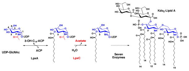
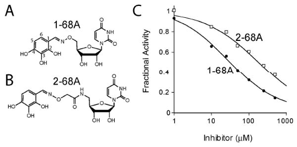
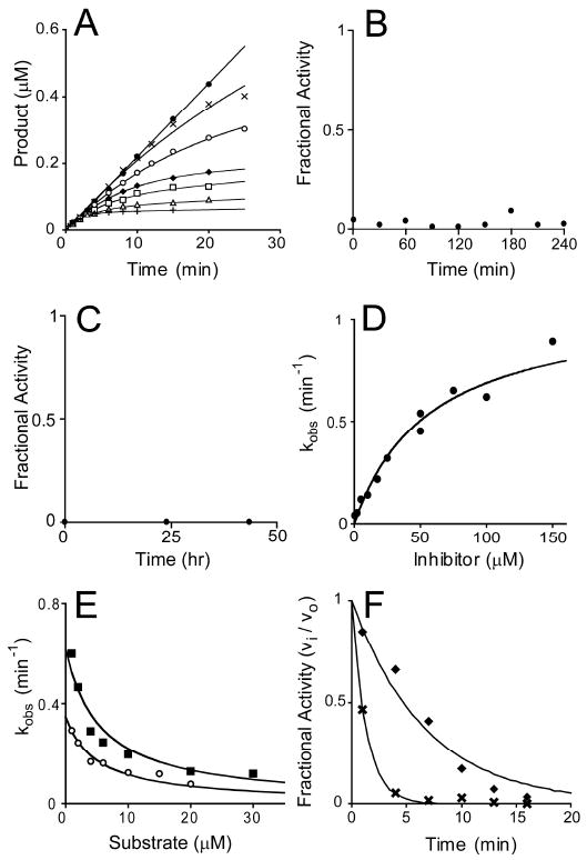
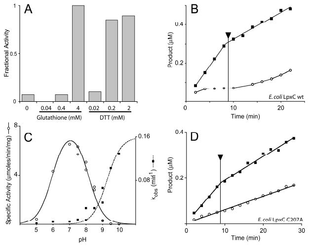
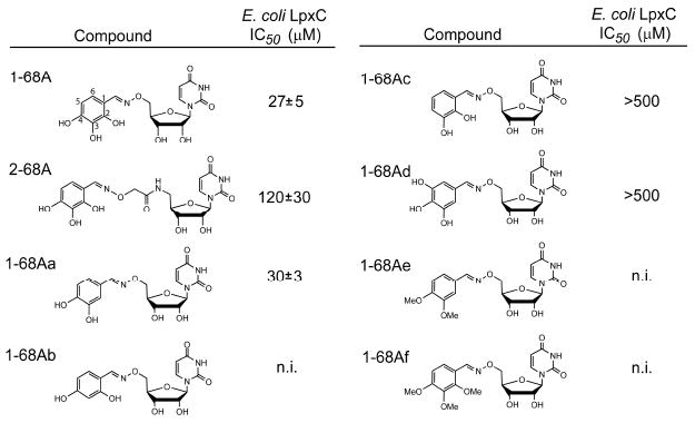
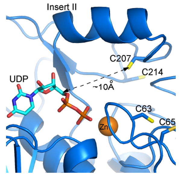
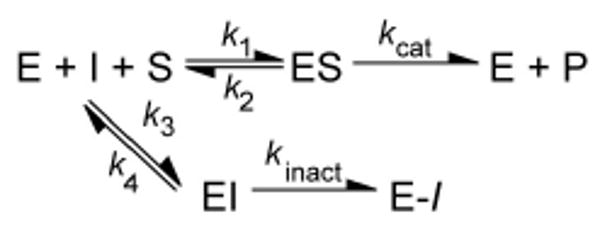
The UDP-3-O-(R-3-hydroxyacyl)-N-acetylglucosamine deacetylase LpxC catalyzes the committed reaction of lipid A (endotoxin) biosynthesis in Gram-negative bacteria and is a validated antibiotic target. Although several previously described compounds bind to the unique acyl chain binding passage of LpxC with high affinity, strategies to target the enzyme's UDP-binding site have not been reported. Here the identification of a series of uridine-based LpxC inhibitors is presented. The most potent examined, 1-68A, is a pH-dependent, two-step, covalent inhibitor of Escherichia coli LpxC that competes with UDP to bind the enzyme in the first step of inhibition. Compound 1-68A exhibits a K(I) of 54 muM and a maximal rate of inactivation (k(inact)) of 1.7 min(-1) at pH 7.4. Dithiothreitol, glutathione and the C207A mutant of E. coli LpxC prevent the formation of a covalent complex by 1-68A, suggesting a role for Cys-207 in inhibition. The inhibitory activity of 1-68A and a panel of synthetic analogues identified moieties necessary for inhibition. 1-68A and a 2-dehydroxy analogue, 1-68Aa, inhibit several purified LpxC orthologues. These compounds may provide new scaffolds for extension of existing LpxC-inhibiting antibiotics to target the UDP binding pocket.


Figures

Figure 1
Figure 1
Reaction catalyzed by LpxC and the structure of E. coli lipid A. The biosynthesis of lipid A begins with the 3-O-acylation of UDP-N-acetylglucosamine by the cytosolic enzyme LpxA (5). In the first irreversible (committed) reaction of the pathway, the deacetylase LpxC unblocks the nitrogen at the 2 position of the glucosamine ring for subsequent acylation by LpxD (5). Seven downstream reactions produce Kdo2-lipid A, the hydrophobic membrane anchor of lipopolysaccharide (5).
Figure 2
Figure 2
Uridine-based inhibitors of E. coli LpxC. Two compounds, 1-68A (Panel A) and 2-68A (Panel B), were identified that inhibited E. coli LpxC with apparent mid- μM IC50s. (Panel C) Analysis of concentration-response plots fitted with Eq. 1 gave IC50 values of 27 μM and 120 μM for 1-68A (black circles) and 2-68A (open squares), respectively, where H=0.7.
Figure 3
Figure 3
Slow, kinetically irreversible and competitive inhibition of E. coli LpxC by 1-68A. (Panel A) Progress curves for product formation when reactions are initiated by the addition of enzyme in the presence 0 μM (black circles), 0.5 μM (black Xs), 2 μM (open circles), 5 μM (black diamonds), 10 μM (open squares), 17.5 μM (open triangles), or 50 μM (black pluses) 1-68A as fitted with Eq. 2. Data is representative of two separate experiments. (Panel B) Rapid dilution of the E. coli LpxC – 1-68A complex. (Panel C) E. coli LpxC activity following dialysis of the LpxC – 1-68A complex. (Panel D) A plot of the observed pseudo first-order rate constant for the formation of the E-I complex (kobs) as fitted with Eq. 3. Repeat experiments (not shown) gave identical results (within 10 %). (Panel E) A plot showing the effect of increasing substrate concentrations on the observed first order rate constant (kobs) in the presence of 10 μM (open circles) or 25 μM (black squares) 1-68A. Eq. 4 was fitted to these data. These plots include data from three independent experiments. (Panel F) The fractional activity of LpxC after a preincubation in the presence of 50 μM 1-68A (black diamonds) with or (black Xs) without 100 mM UDP. The fit of a first-order, decaying exponential (vi / vo = e-kt) for each data set is shown as a line.
Figure 4
Figure 4
Inhibition of E. coli LpxC by 1-68A depends upon a thiol group. (Panel A) Effect of glutathione or dithiothreitol (DTT) on 1-68A inhibition of E. coli LpxC activity. (Panel B) Reactions containing 0 μM (black squares) or 50 μM (open circles) 1-68A were initiated with 0.1 nM E. coli LpxC. Both reactions progressed for 9 min, at which point dithiothreitol was added (as indicated by the arrow) to a final concentration of 2 mM. The reduced velocity of the 0 μM reaction after 9 min is not due to dilution but rather dithiothreitol is a weak competitive inhibitor E. coli LpxC (data not shown). (Panel C) pH rate profile of wild-type E. coli LpxC specific activity (open circles) and the rate (kobs) of 1-68A inhibition (black squares). The bell-shaped profile is fit with a pK1 of 5.9 and a pK2 of 8.0. The kobs profile is fitted with a curve describing a pKa of at least 9.2. (Panel D) Reactions containing 0 μM (black squares) or 2 mM (open circles) 1-68A were initiated with 0.1 nM E. coli LpxC C207A. Both reactions progressed for 9 min, at which point dithiothreitol was added (as indicated by the arrow) to a final concentration of 2 mM. Data is representative of two separate experiments.
Figure 5
Figure 5
ESI-MS analysis of the E. coli LpxC – 1-68A complex. (Panel A) The LC-elution profile of E. coli LpxC preincubated with equimolar 1-68A, shown as the extracted ion current. Positive ion (Panel B) and deconvoluted (Panel C) mass spectra of E. coli LpxC from the 16-22 min region of the LC elution shown in Panel A. The observed mass of the E. coli LpxC polypeptide was 33,952 Da (data not shown). (Panel D) Mass spectrum of products formed after incubating N-acetylcysteine-methylester and 1-68A at pH 7.4. Note: The proposed structure of b, i.e. covalent complex of N-acetylcysteine-methylester and 1-68A, was proposed based on exact mass measurement and MS/MS, which would not delineate position isomers. (Panel E) MS/MS analysis of the “b” ion in Panel D.
Figure 6
Figure 6. E. coli
LpxC inhibition by 1-68A analogs. Analogs of 1-68A containing variable substituents on the benzene ring were synthesized and tested for inhibition of E. coli LpxC. Of this series, only 1-68Aa was of comparable potency to 1-68A, and is likewise a time-dependent inhibitor of E. coli and Helicobacter pylori LpxC (data not shown).
Figure 7
Figure 7
Potential binding mode of UDP and location of cysteine residues in the E. coli LpxC active site. This model is based upon an A. aeolicus LpxC – UDP complex, as determined by x-ray crystallography (2IER). The zinc ion is shown as an orange sphere. This figure was prepared using PyMOL.
Scheme 1
Scheme 1
Mechanism for two-step, kinetically irreversible inhibition.

Similar articles

Cited by

References

    1. Walsh C. Molecular mechanisms that confer antibacterial drug resistance. Nature. 2000;406:775–781. - PubMed
    1. Projan SJ, Youngman PJ. Antimicrobials: new solutions badly needed. Curr Opin Microbiol. 2002;5:463–465. - PubMed
    1. Russell AD, Chopra I. Understanding antibacterial action and resistance. E. Horwood; New York: 1990.
    1. Wyckoff TJ, Raetz CRH, Jackman JE. Antibacterial and anti-inflammatory agents that target endotoxin. Trends Microbiol. 1998;6:154–159. - PubMed
    1. Raetz CRH, Whitfield C. Lipopolysaccharide endotoxins. Annu Rev Biochem. 2002;71:635–700. - PMC - PubMed

Publication types

MeSH terms