

Sustained BMP signaling in osteoblasts stimulates bone formation by promoting angiogenesis and osteoblast differentiation
- PMID: 19257813
- PMCID: PMC2697625
- DOI: 10.1359/jbmr.090204
Sustained BMP signaling in osteoblasts stimulates bone formation by promoting angiogenesis and osteoblast differentiation
Abstract
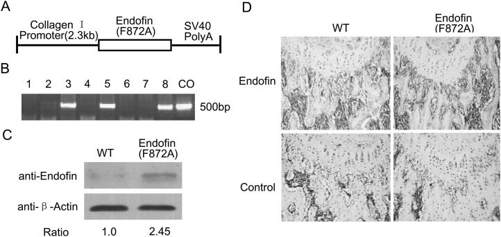
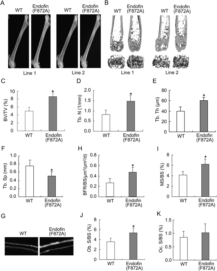
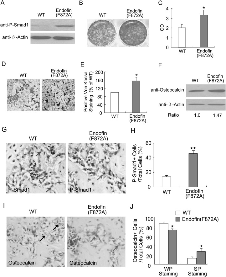
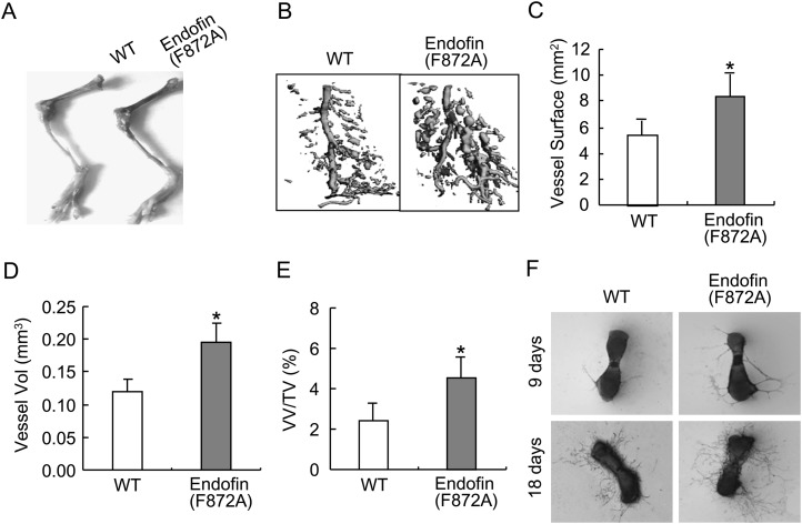
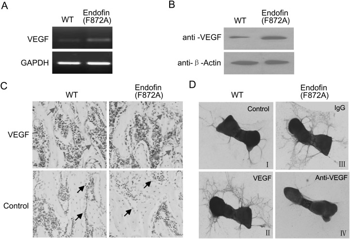
Angiogenesis and bone formation are tightly coupled during the formation of the skeleton. Bone morphogenetic protein (BMP) signaling is required for both bone development and angiogenesis. We recently identified endosome-associated FYVE-domain protein (endofin) as a Smad anchor for BMP receptor activation. Endofin contains a protein-phosphatase pp1c binding domain, which negatively modulates BMP signals through dephosphorylation of the BMP type I receptor. A single point mutation of endofin (F872A) disrupts interaction between the catalytic subunit pp1c and sensitizes BMP signaling in vitro. To study the functional impact of this mutation in vivo, we targeted expression of an endofin (F872A) transgene to osteoblasts. Mice expressing this mutant transgene had increased levels of phosphorylated Smad1 in osteoblasts and showed increased bone formation. Trabecular bone volume was significantly increased in the transgenic mice compared with the wildtype littermates with corresponding increases in trabecular bone thickness and number. Interestingly, the transgenic mice also had a pronounced increase in the density of the bone vasculature measured using contrast-enhanced microCT imaging of Microfil-perfused bones. The vessel surface and volume were both increased in association with elevated levels of vascular endothelial growth factor (VEGF) in osteoblasts. Endothelial sprouting from the endofin (F872A) mutant embryonic metatarsals cultured ex vivo was increased compared with controls and was abolished by an addition of a VEGF neutralizing antibody. In conclusion, osteoblast targeted expression of a mutant endofin protein lacking the pp1c binding activity results in sustained signaling of the BMP type I receptor, which increases bone formation and skeletal angiogenesis.
Figures
Similar articles
-
Endofin acts as a Smad anchor for receptor activation in BMP signaling.J Cell Sci. 2007 Apr 1;120(Pt 7):1216-24. doi: 10.1242/jcs.03400. Epub 2007 Mar 13. J Cell Sci. 2007. PMID: 17356069
-
Role of bone morphogenetic proteins in sprouting angiogenesis: differential BMP receptor-dependent signaling pathways balance stalk vs. tip cell competence.FASEB J. 2017 Nov;31(11):4720-4733. doi: 10.1096/fj.201700193RR. Epub 2017 Jul 21. FASEB J. 2017. PMID: 28733457 Free PMC article.
-
Controversy of physiological vs. pharmacological effects of BMP signaling: Constitutive activation of BMP type IA receptor-dependent signaling in osteoblast lineage enhances bone formation and resorption, not affecting net bone mass.Bone. 2020 Sep;138:115513. doi: 10.1016/j.bone.2020.115513. Epub 2020 Jun 27. Bone. 2020. PMID: 32603910 Free PMC article.
-
Bone morphogenetic proteins.Growth Factors. 2004 Dec;22(4):233-41. doi: 10.1080/08977190412331279890. Growth Factors. 2004. PMID: 15621726 Review.
-
TGF-β and BMP signaling in osteoblast differentiation and bone formation.Int J Biol Sci. 2012;8(2):272-88. doi: 10.7150/ijbs.2929. Epub 2012 Jan 21. Int J Biol Sci. 2012. PMID: 22298955 Free PMC article. Review.
Cited by
-
Novel candidate genes putatively involved in stress fracture predisposition detected by whole-exome sequencing.Genet Res (Camb). 2014;96:e004. doi: 10.1017/S001667231400007X. Genet Res (Camb). 2014. PMID: 25023003 Free PMC article.
-
Osteoinductivity Assessment of BMP-2 Loaded Composite Chitosan-Nano-Hydroxyapatite Scaffolds in a Rat Muscle Pouch.Materials (Basel). 2011 Aug 2;4(8):1360-1374. doi: 10.3390/ma4081360. Materials (Basel). 2011. PMID: 28824147 Free PMC article.
-
Genomic analyses of early peri-implant bone healing in humans: a systematic review.Int J Implant Dent. 2015 Dec;1(1):5. doi: 10.1186/s40729-015-0006-2. Epub 2015 Mar 1. Int J Implant Dent. 2015. PMID: 27747627 Free PMC article. Review.
-
Bone morphogenetic protein 2 stimulates endochondral ossification by regulating periosteal cell fate during bone repair.Bone. 2010 Jul;47(1):65-73. doi: 10.1016/j.bone.2010.03.012. Epub 2010 Mar 27. Bone. 2010. PMID: 20348041 Free PMC article.
-
In vitro model of vascularized bone: synergizing vascular development and osteogenesis.PLoS One. 2011;6(12):e28352. doi: 10.1371/journal.pone.0028352. Epub 2011 Dec 2. PLoS One. 2011. PMID: 22164277 Free PMC article.
References
-
- Adams SL, Cohen AJ, Lassová L. Integration of signaling pathways regulating chondrocyte differentiation during endochondral bone formation. J Cell Physiol. 2007;213:635–641. - PubMed
-
- Gerber HP, Ferrara N. Angiogenesis and bone growth. Trends Cardiovasc Med. 2000;10:223–228. - PubMed
-
- Carano RA, Filvaroff EH. Angiogenesis and bone repair. Drug Discov Today. 2003;8:980–989. - PubMed
-
- Coultas L, Chawengsaksophak K, Rossant J. Endothelial cells and VEGF in vascular development. Nature. 2005;438:937–945. - PubMed
-
- Chen D, Ji X, Harris MA, Feng JQ, Karsenty G, Celeste AJ, Rosen V, Mundy GR, Harris SE. Differential roles for bone morphogenetic protein (BMP) receptor type IB and IA in differentiation and specification of mesenchymal precursor cells to osteoblast and adipocyte lineages. J Cell Biol. 1998;142:295–305. - PMC - PubMed
Publication types
MeSH terms
Substances
Grants and funding
LinkOut - more resources
Full Text Sources
Molecular Biology Databases
