. 2009 Aug;24(8):1481-92.
doi: 10.1359/jbmr.090226.

Serum IGF-1 determines skeletal strength by regulating subperiosteal expansion and trait interactions

Affiliations

Serum IGF-1 determines skeletal strength by regulating subperiosteal expansion and trait interactions

Shoshana Yakar et al. J Bone Miner Res. 2009 Aug.

Abstract

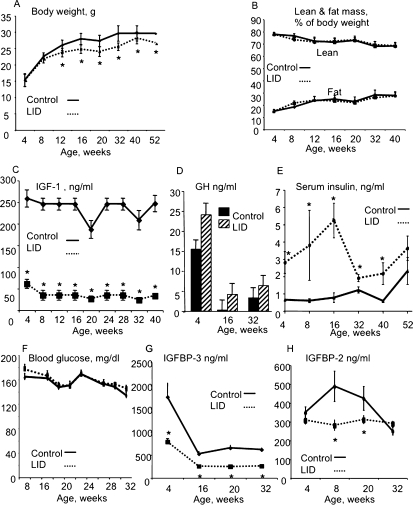
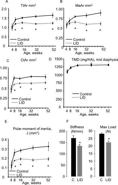
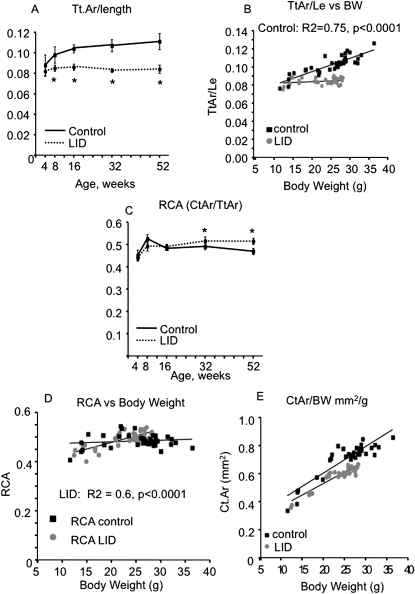
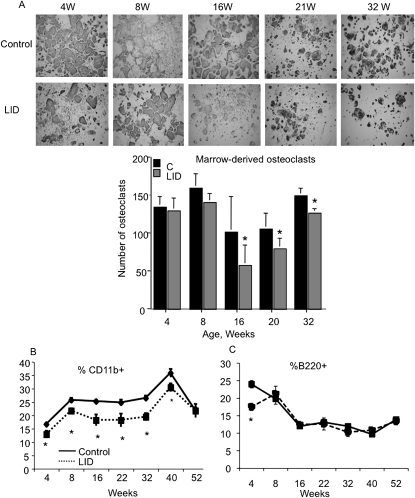
Strong correlations between serum IGF-1 levels and fracture risk indicate that IGF-1 plays a critical role in regulating bone strength. However, the mechanism by which serum IGF-1 regulates bone structure and fracture resistance remains obscure and cannot be determined using conventional approaches. Previous analysis of adult liver-specific IGF-1-deficient (LID) mice, which exhibit 75% reductions in serum IGF-1 levels, showed reductions in periosteal circumference, femoral cross-sectional area, cortical thickness, and total volumetric BMD. Understanding the developmental sequences and the resultant anatomical changes that led to this adult phenotype is the key for understanding the complex relationship between serum IGF-1 levels and fracture risk. Here, we identified a unique developmental pattern of morphological and compositional traits that contribute to bone strength. We show that reduced bone strength associated with low levels of IGF-1 in serum (LID mice) result in impaired subperiosteal expansion combined with impaired endosteal apposition and lack of compensatory changes in mineralization throughout growth and aging. We show that serum IGF-1 affects cellular activity differently depending on the cortical surface. Last, we show that chronic reductions in serum IGF-1 indirectly affect bone strength through its effect on the marrow myeloid progenitor cell population. We conclude that serum IGF-1 not only regulates bone size, shape, and composition during ontogeny, but it plays a more fundamental role-that of regulating an individual's ability to adapt its bone structure to mechanical loads during growth and development.


Figures

FIG. 1
FIG. 1
Control and LID mice were followed from 4 to 52 wk of age. Body weight (A) and body composition (B) were followed monthly by MRI. Serum was obtained through the mandibular vein once a month, and IGF-1 (C), GH (D), insulin (E), blood glucose (F), IGFBP-3 (G), and IGFBP-2 (H) levels were analyzed. Results are presented as mean ± SE of at least 40 mice per age group per genotype.
FIG. 2
FIG. 2
Trabecular bone fraction does not differ significantly between control and LID mice. Trabecular bone parameters were assessed by μCT, and BV/TV (A) and TMD (B) were measured at the distal femur of formalin-fixed bones. Histomorphometry was performed on plastic bone sections from the distal femur; %Er.Bs (C), %OCs/Bs (D), N.OC/Ta (E), and %Os/Bs (F) were determined at 4, 8, and 20 wk of age. Results are presented as mean ± SE of n = 6 mice per age group per genotype.
FIG. 3
FIG. 3
LID mice have slender and more fragile bones. Cortical bone parameters were assessed during growth at the femoral midshaft using μCT. LID mice show reduced Tt.Ar. (A), Ma.Ar (B), and Ct.Ar (C) starting at 8 wk of age. TMD (D), measured by μCT, did not differ significantly between control and LID mice at all ages. LID mice cannot restore mechanical properties; LID mice show decreased polar moment of inertia (E) throughout growth and reduced stiffness and max load at four-point bending assay at 20 wk of age (F). Results are presented as mean ± SD of n > 8 mice per age group per genotype.
FIG. 4
FIG. 4
LID mice exhibit impaired co-variation of morphological traits (obtained by μCT). LID mice have slender bones throughout growth starting at 4 wk (Tt.Ar/length) (A). Control mice show a positive relationship between Tt.Ar/length and body weight, whereas LID mice do not (B). LID mice exhibit an increase in relative cortical area, RCA, at 32 and 52 wk of age (C). LID mice show a positive relationship between RCA and body weight (D), suggesting that these mice compensate for the slender bones by increasing cortical area. Both control and LID mice show positive relationship between Ct.Ar and body weight (E). Results are presented as mean ± SD of n > 8 mice age per group per genotype.
FIG. 5
FIG. 5
The number of osteoclast precursors in the nonadherent population derived from LID mice is decreased. Osteoclastogenesis was induced in nonadherent cells derived from marrow of LID and control mice (A). TRACP+ cells were detected after 5–6 days in culture; the bar graph represents quantification of osteoclast number in cultures. FACS analysis of cell surface markers in marrow derived from control and LID mice. Percent of CD11b+ cells was reduced in LID mice throughout development (B), whereas percent B220+ cells reduced significantly only at 4 wk of age. Results are presented as mean ± SE of n > 8 mice age per group per genotype.
FIG. 6
FIG. 6
Schematic representation of the cortical bone envelope during growth. Control mice show a marked increase in cross-sectional area during the first 8 wk of life and later on a slight increase in cross-sectional area associated with expansion of marrow area to allow an efficient structure to support the age-related increase in body weight. LID mice, however, show a marked increase in cross-sectional area during the first 8 wk of life but have a thinner cortical envelope. Later in life, LID mice fail to increase cross-sectional area (because of impaired subperiosteal expansion) and instead show endosteal apposition in an effort to support the age-related increase in body weight.

Similar articles

Cited by

References

    1. Langlois JA, Rosen CJ, Visser M, Hannan MT, Harris T, Wilson PW, Kiel DP. Association between insulin-like growth factor I and bone mineral density in older women and men: The Framingham Heart Study. J Clin Endocrinol Metab. 1998;83:4257–4262. - PubMed
    1. Garnero P, Sornay-Rendu E, Delmas PD. Low serum IGF-1 and occurrence of osteoporotic fractures in postmenopausal women. Lancet. 2000;355:898–899. - PubMed
    1. Turner CH. Functional determinants of bone structure: Beyond Wolff's law of bone transformation. Bone. 1992;13:403–409. - PubMed
    1. Beck TJ, Ruff CB, Mourtada FA, Shaffer RA, Maxwell-Williams K, Kao GL, Sartoris DJ, Brodine S. Dual-energy X-ray absorptiometry derived structural geometry for stress fracture prediction in male U.S. Marine Corps recruits. J Bone Miner Res. 1996;11:645–653. - PubMed
    1. Black DM, Bouxsein ML, Marshall LM, Cummings SR, Lang TF, Cauley JA, Ensrud KE, Nielson CM, Orwoll ES. Proximal femoral structure and the prediction of hip fracture in men: A large prospective study using QCT. J Bone Miner Res. 2008;23:1326–1333. - PMC - PubMed

Publication types

Substances