

Functional characterization of E- and P-cadherin in invasive breast cancer cells
- PMID: 19257890
- PMCID: PMC2656544
- DOI: 10.1186/1471-2407-9-74
Functional characterization of E- and P-cadherin in invasive breast cancer cells
Abstract
Background: Alterations in the cadherin-catenin adhesion complexes are involved in tumor initiation, progression and metastasis. However, the functional implication of distinct cadherin types in breast cancer biology is still poorly understood.
Methods: To compare the functional role of E-cadherin and P-cadherin in invasive breast cancer, we stably transfected these molecules into the MDA-MB-231 cell line, and investigated their effects on motility, invasion and gene expression regulation.
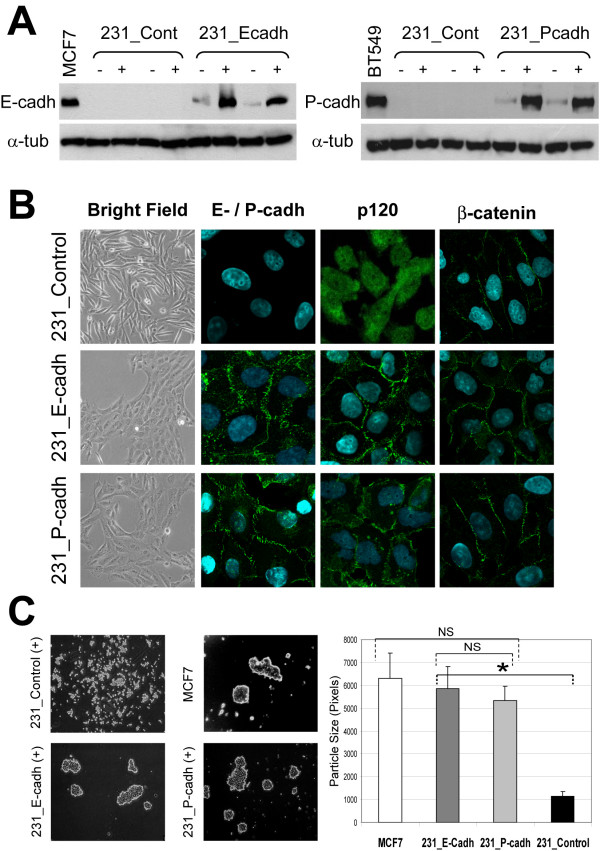
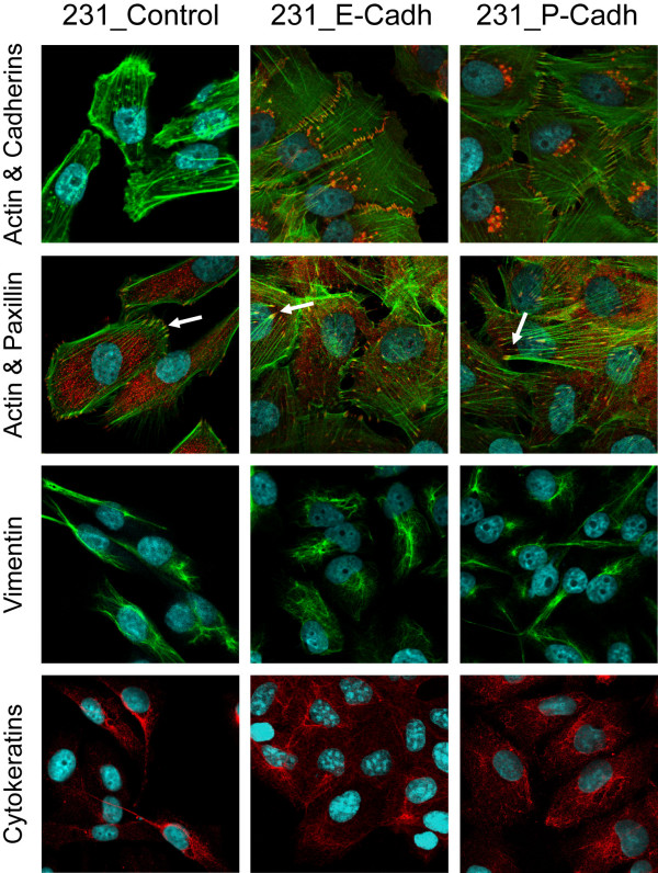
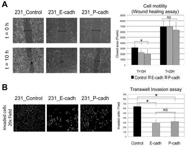
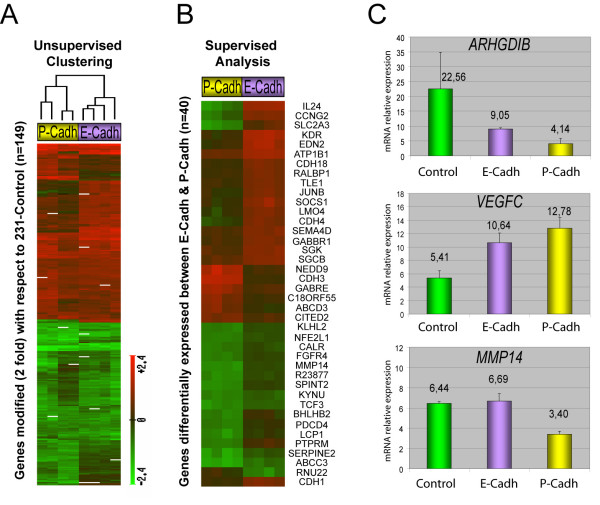
Results: Expression of either E- and P-cadherin significantly increased cell aggregation and induced a switch from fibroblastic to epithelial morphology. Although expression of these cadherins did not completely reverse the mesenchymal phenotype of MDA-MB-231 cells, both E- and P-cadherin decreased fibroblast-like migration and invasion through extracellular matrix in a similar way. Moreover, microarray gene expression analysis of MDA-MB-231 cells after expression of E- and P-cadherins revealed that these molecules can activate signaling pathways leading to significant changes in gene expression. Although the expression patterns induced by E- and P-cadherin showed more similarities than differences, 40 genes were differentially modified by the expression of either cadherin type.
Conclusion: E- and P-cadherin have similar functional consequences on the phenotype and invasive behavior of MDA-MB-231 cells. Moreover, we demonstrate for the first time that these cadherins can induce both common and specific gene expression programs on invasive breast cancer cells. Importantly, these identified genes are potential targets for future studies on the functional consequences of altered cadherin expression in human breast cancer.
Figures
Similar articles
-
Epithelial requirement for in vitro proliferation and xenograft growth and metastasis of MDA-MB-468 human breast cancer cells: oncogenic rather than tumor-suppressive role of E-cadherin.Breast Cancer Res. 2017 Jul 27;19(1):86. doi: 10.1186/s13058-017-0880-z. Breast Cancer Res. 2017. PMID: 28750639 Free PMC article.
-
GATA3 inhibits breast cancer metastasis through the reversal of epithelial-mesenchymal transition.J Biol Chem. 2010 Apr 30;285(18):14042-51. doi: 10.1074/jbc.M110.105262. Epub 2010 Feb 26. J Biol Chem. 2010. PMID: 20189993 Free PMC article.
-
E-cadherin expression in human breast cancer cells suppresses the development of osteolytic bone metastases in an experimental metastasis model.Cancer Res. 1996 Sep 1;56(17):4063-70. Cancer Res. 1996. PMID: 8752180
-
The role of E-cadherin and scatter factor in tumor invasion and cell motility.EXS. 1991;59:109-26. doi: 10.1007/978-3-0348-7494-6_8. EXS. 1991. PMID: 1833225 Review.
-
P-cadherin role in normal breast development and cancer.Int J Dev Biol. 2011;55(7-9):811-22. doi: 10.1387/ijdb.113382aa. Int J Dev Biol. 2011. PMID: 22161837 Review.
Cited by
-
Active wetting of epithelial tissues.Nat Phys. 2019 Jan;15(1):79-88. doi: 10.1038/s41567-018-0279-5. Epub 2018 Sep 24. Nat Phys. 2019. PMID: 31537984 Free PMC article.
-
Down-regulation of the oncogene cyclin D1 increases migratory capacity in breast cancer and is linked to unfavorable prognostic features.Am J Pathol. 2010 Dec;177(6):2886-97. doi: 10.2353/ajpath.2010.100303. Epub 2010 Oct 22. Am J Pathol. 2010. PMID: 20971731 Free PMC article.
-
Macro- and micro-nutrient-based multiplex stress conditions modulate in vitro tumorigenesis and aggressive behavior of breast cancer spheroids.In Vitro Model. 2021 Nov 25;1(1):85-101. doi: 10.1007/s44164-021-00006-5. eCollection 2022 Feb. In Vitro Model. 2021. PMID: 39872971 Free PMC article.
-
p120ctn and P-cadherin but not E-cadherin regulate cell motility and invasion of DU145 prostate cancer cells.PLoS One. 2010 Jul 27;5(7):e11801. doi: 10.1371/journal.pone.0011801. PLoS One. 2010. PMID: 20668551 Free PMC article.
-
Brain metastases from lung cancer show increased expression of DVL1, DVL3 and beta-catenin and down-regulation of E-cadherin.Int J Mol Sci. 2014 Jun 13;15(6):10635-51. doi: 10.3390/ijms150610635. Int J Mol Sci. 2014. PMID: 24933634 Free PMC article.
References
Publication types
MeSH terms
Substances
LinkOut - more resources
Full Text Sources
Medical
Molecular Biology Databases
Miscellaneous
