. 2009 Mar 3:9:15.
doi: 10.1186/1472-6750-9-15.

Addition of a sequence from alpha2-antiplasmin transforms human serum albumin into a blood clot component that speeds clot lysis

Affiliations

Addition of a sequence from alpha2-antiplasmin transforms human serum albumin into a blood clot component that speeds clot lysis

William P Sheffield et al. BMC Biotechnol. .

Abstract

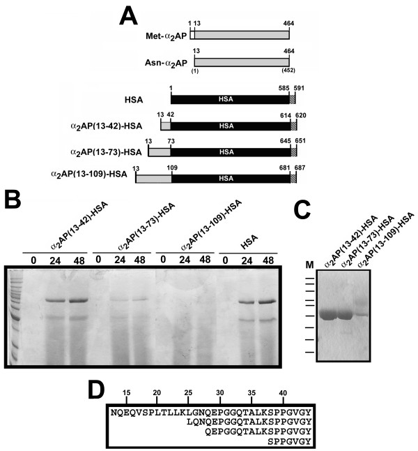
Background: The plasma protein alpha2-antiplasmin (alpha2AP) is cross-linked to fibrin in blood clots by the transglutaminase factor XIIIa, and in that location retards clot lysis. Competition for this effect could be clinically useful in patients with thrombosis. We hypothesized that fusion of N-terminal portions of alpha2-antiplasmin to human serum albumin (HSA) and production of the chimeric proteins in Pichia pastoris yeast would produce a stable and effective competitor protein.

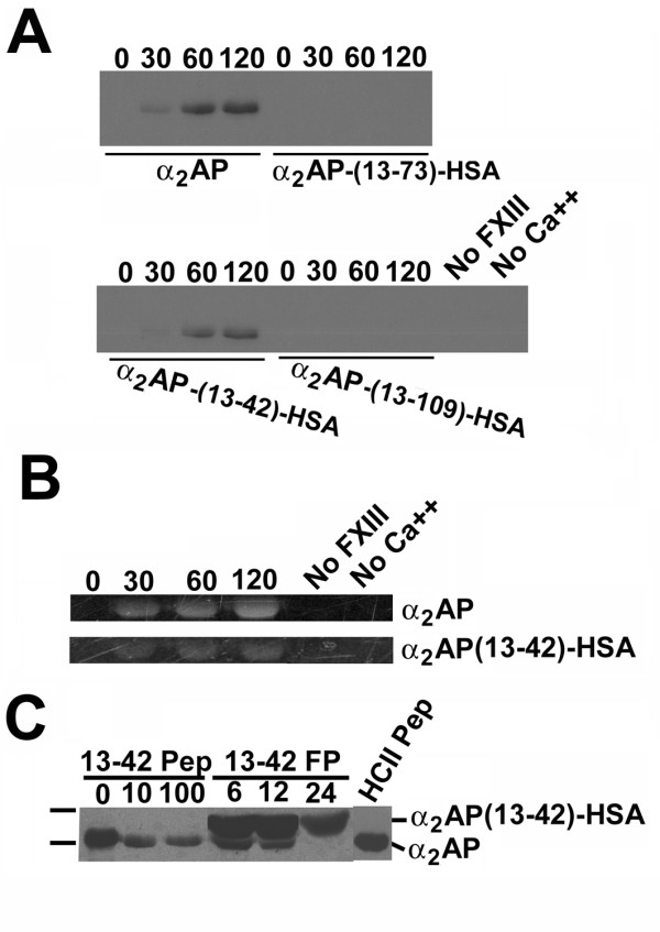
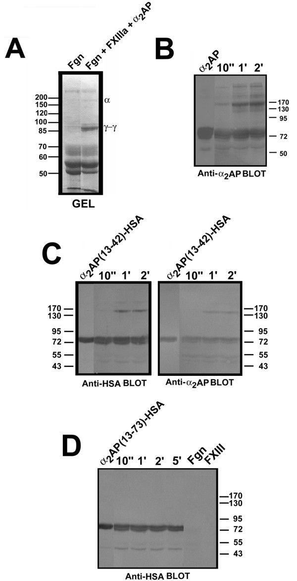
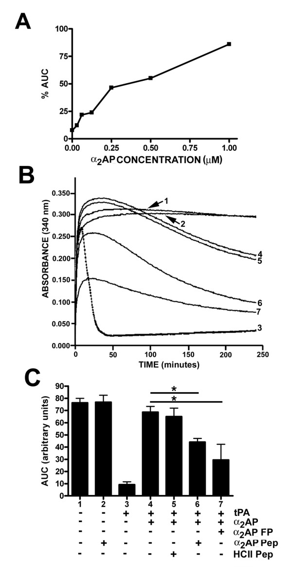
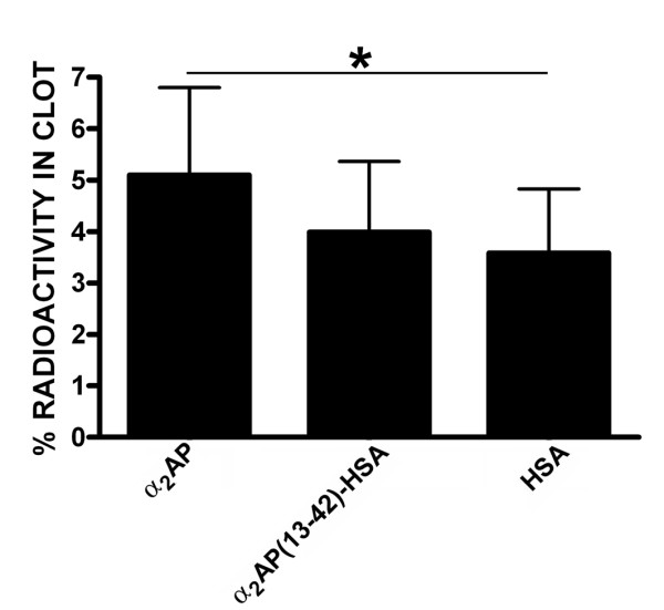
Results: Fusion protein alpha2AP(13-42)-HSA was efficiently secreted from transformed yeast and purified preparations contained within a mixed population the full-length intact form, while fusions with longer alpha2AP moieties were inefficiently secreted and/or degraded. The alpha2AP(13-42)-HSA protein, but not recombinant HSA, was cross-linked to both chemical lysine donors and fibrin or fibrinogen by factor XIIIa, although with less rapid kinetics than native alpha2AP. Excess alpha2AP(13-42)-HSA competed with alpha2AP for cross-linking to chemical lysine donors more effectively than a synthetic alpha2AP(13-42) peptide, and reduced the alpha2AP-dependent resistance to fibrinolysis of plasma clots equally effectively as the peptide. Native alpha2AP was found in in vivo clots in rabbits to a greater extent than alpha2AP(13-42), however.

Conclusion: In this first report of transfer of transglutamination substrate status from one plasma protein to another, fusion protein alpha2AP(13-42)-HSA was shown to satisfy initial requirements for a long-lasting, well-tolerated competitive inhibitor of alpha2-antiplasmin predicted to act in a clot-localized manner.


Figures

Figure 1
Figure 1
α2AP-HSA fusion protein design, expression, and characterization. (A) Schematic diagram of proteins. Relevant polypeptides are shown in linear form, with important residues identified above each bar. White bars correspond to the N-terminal dodecapeptide sequence removed from Met-α2AP to form Asn-α2AP, shown in grey; HSA is shown in black, and C-terminal hexahistidines are shown as stippled bars. (B) Electrophoretic profile of conditioned media samples taken from P. pastoris cultures at the times indicated above the lanes, post-induction with methanol. A Coomassie Blue-stained 10% SDS-polyacrylamide gel is shown. Cell lines were transformed with plasmid constructs directing the synthesis of the proteins identified above the horizontal lines. Markers on the leftmost lane of the gel are, in kDa: 160; 140; 120; 100; 90; 80; 70; 60; 50; 40; 30; and 25. (C) A stained gel similar that in panel B is shown, except that 5 μg of the purified proteins identified above the lanes were electrophoresed. M, markers, same as in panel B. (D) N-termini found in purified α2AP(13-42)-HSA by amino acid sequencing of the preparation shown in the leftmost lane of panel C. Numbers above the box identify every fifth amino acid residue in α2AP(13-42)-HSA.
Figure 2
Figure 2
Factor XIIIa-mediated incorporation of BPA or dansyl cadaverine into α2AP and fusion proteins. (A) Relevant portions of streptavidin blots containing the reaction products of the proteins identified below the panels with 10 mM BPA in the presence of thrombin, FXIII, calcium, and 7.14 μM test proteins, for the times in minutes shown above the lanes, using reaction conditions described in "Methods". Lanes labelled "No FXIII" and "No Ca++" correspond to reactions identical to those shown for α2AP at 120 minutes, with the omission of the listed components. (B) Portions of 8% SDS-acrylamide gels visualized by ultraviolet transillumination following fXIIIa-mediated transglutamination of 0.5 mM dansyl cadaverine to the proteins listed the right of the panels. (C) Same as panel A, except that all reactions contained α2AP at 1.7 μM, reacted with BPA, thrombin and FXIII in the presence of the concentrations of either α2AP(13-42) synthetic peptide (13-42 Pep), α2AP(13-42)-HSA (13-42 FP for fusion protein) shown above the lanes. The positions of α2AP and α2AP(13-42)-HSA are highlighted at right.
Figure 3
Figure 3
Cross-linking of α2AP and α2AP(13-42)-HSA by fXIIIa to fibrinogen. FXIII was pre-activated to fXIIIa by thrombin, the thrombin inactivated with FPRck, and the fXIIIa then combined with fibrinogen and α2AP or α2AP(13-42)-HSA or α2AP(13-73)-HSA in transglutamination reactions. Reactions were terminated with SDS, DTT, and urea as described in "Methods". (A) depicts a Coomassie Blue-stained SDS-polyacrylamide gel highlighting the cross-linking of fibrinogen into γ-γ dimers (γ-γ) and α-polymers (α). (B) shows an anti-α2AP immunoblot of transglutamination reactions containing α2AP for the times identified above the lanes; the first lane contains α2AP alone. (C), right panel, is identical to B except that α2AP(13-42)-HSA was substituted for α2AP; (C), left panel, is identical to the right panel except an anti-HSA antibody was used. (D) is identical to (C), left panel, except that α2AP(13-73)-HSA was substituted for α2AP(13-42)-HSA. The position of molecular mass markers, is shown to the left or right of the panels.
Figure 4
Figure 4
Effects of α2AP and derivatives on plasma clot formation and lysis. (A) Clot formation and lysis was followed by monitoring turbidity (absorbance at 340 nm) every 30 seconds for 4 hours using a plate reader, of clots formed using diluted α2AP-deficient plasma containing both 5 nM thrombin and 0.125 nM tPA, and taking the area under the turbidity versus time curve (AUC). Reactions were supplemented with increasing concentrations of purified plasma-derived α2AP, and the AUC relative to that of reactions lacking tPA reported as a percentage. Results of a single experiment are shown. (B) shows turbidity plots for reactions similar to those shown in A, under 7 conditions described in the + or - table in panel C; for instance, (1) shows stable clot formation in the absence of tPA, (3) shows clot formation and rapid lysis in the presence of tPA, (4) shows attenuation of clot lysis in the presence of 1.0 μM α2AP and (6) and (7) show competition of the α2AP effect by 14 μM α2AP(13-42) synthetic peptide (Pep) or α2AP(13-42)-HSA fusion protein (FP), respectively. C shows the results of quantification of the experiment shown in B and repeated 4 times (n = 3 to 11 ± SD), under the conditions summarized below the graph, as indicated below the lanes. A control peptide unrelated to α2AP corresponding to residues 54–75 of heparin cofactor II was used at 14 μM (HCII Pep). Asterisks indicate significant differences between groups compared between the horizontal lines (p < 0.05).
Figure 5
Figure 5
Localization of radiolabeled proteins in rabbit jugular vein clots in vivo. The radioactivity remaining in rabbit jugular vein clots allowed to polymerize in clamped-off vessels in situ for 30 minutes in the anesthetized animal, then to age with circulation restored for 60 minutes, prior to clot recovery and γ-counting is shown. Clots formed in the presence of 125I-fibrinogen and 131I-labelled plasma-derived α2AP or recombinant α2AP(13-42)-HSA or recombinant HSA. Individual data points are the mean of values for both left and right jugular veins, shown as the mean of 6 such means ± SD). Asterisk indicates the only comparison in the group significant (p < 0.05) by paired t-tests.

Similar articles

Cited by

References

    1. Bornberg-Bauer E, Beaussart F, Kummerfeld SK, Teichmann SA, Weiner J., 3rd The evolution of domain arrangements in proteins and interaction networks. Cell Mol Life Sci. 2005;62:435–445. - PMC - PubMed
    1. Koonin EV. How many genes can make a cell: the minimal-gene-set concept. Annu Rev Genomics Hum Genet. 2000;1:99–116. - PMC - PubMed
    1. Sheffield WP. Modification of clearance of therapeutic and potentially therapeutic proteins. Curr Drug Targets Cardiovasc Haematol Disord. 2001;1:1–22. - PubMed
    1. Peters T., Jr Serum albumin. Adv Protein Chem. 1985;37:161–245. - PubMed
    1. Dugaiczyk A, Law SW, Dennison OE. Nucleotide sequence and the encoded amino acids of human serum albumin mRNA. Proc Natl Acad Sci USA. 1982;79:71–75. - PMC - PubMed

Publication types

LinkOut - more resources