. 2009 May;141(2-3):214-21.
doi: 10.1016/j.bpc.2009.02.001. Epub 2009 Feb 12.

Kinetic folding studies of the P22 tailspike beta-helix domain reveal multiple unfolded states

Affiliations

Kinetic folding studies of the P22 tailspike beta-helix domain reveal multiple unfolded states

M L Spatara et al. Biophys Chem. 2009 May.

Abstract

The beta-helix is an important protein fold in many pathogens, and is a challenging system for folding pathway prediction because it primarily is stabilized by non-local interactions along the primary sequence. A useful experimental model of this fold is a monomeric truncation of P22 tailspike protein, the beta-helix domain (bhx). This report describes a systematic in vitro study of the chemical denaturation and refolding of bhx. Results from equilibrium chemical denaturation experiments were consistent with a two-state folding mechanism, but showed only partial reversibility. Stopped-flow fluorescence studies showed a single unfolding step, but two refolding steps. The slow refolding step could be partly attributed to proline isomerization, based on an increased rate during refolding in the presence of PPIase and an increased relative amplitude of this step with increasing delay time in double-jump refolding experiments observed over delays of 5-100 s. However, double-jump refolding experiments with delay times longer than 100 s along with size exclusion chromatography and dynamic light scattering of refolding samples showed that the overall refolding yield decreased as bhx was unfolded for longer periods of time. Furthermore, the losses resulted from aggregate formation during refolding. This suggests that a change occurs over time in the unfolded or denatured state ensemble that increases the propensity for aggregation upon the shift to more native-favoring conditions. Alternatively aggregate nuclei may be able to form even under high denaturant conditions, and these subsequently grow and consume monomer when placed under native-favoring conditions.


Figures

Fig. 1.
Fig. 1.
P22 tailspike protein structure. Ribbon diagram of full-length, trimeric tailspike protein. Arrows indicate the approximate locations of residues 109 and 544, the truncation points for bhx.
Fig. 2.
Fig. 2.
Equilibrium folding of bhx. (a) Intrinsic fluorescence spectra of bhx in 0 M (_) and 6 M (…) urea. Denatured protein shows a decrease in intensity along with a small red shift. Spectral center of mass (b) and emission intensity at 340 nm (c) are shown for bhx unfolding (○) and refolding (●) as a function of final urea concentration.
Fig. 3.
Fig. 3.
Kinetics of (un)folding. Representative kinetic traces for unfolding (a) and refolding (b), (c) of bhx. Experimental data (○) and fits to a summed exponential expression (_) with one (a), (b) or two (c) terms are shown in the main plot. Residuals for the shown fit are plotted below. For clarity, plots show every third experimental point.
Fig. 4.
Fig. 4.
Chevron plot representation of kinetic folding data. The urea dependence of the rate constants for unfolding (○) as well as the slow (■) and fast (▼) folding phases of bhx. Error bars represent the standard deviation from at least three experiments, and are smaller than the size of the symbol where not shown.
Fig. 5.
Fig. 5.
Effect of PPIase. Refolding kinetic traces for bhx with (♦) and without (+) PPIase.
Fig. 6.
Fig. 6.
Sequential-mixing refolding. Samples of bhx were allowed to unfold in high concentration urea solutions for varying delay times, before being refolded by dilution into low urea conditions. The rate constants (a,c) and relative amplitudes (b,d) of the fast (▼) and slow (■) refolding phases of bhx are plotted as a function of the refolding delay time. Error bars for data from long delay times (c,d) represent the 95% confidence intervals from the non-linear regression. For data with short delay times, where variability between runs was significant, error bars represent the standard deviation in the parameters from fits of at least 5 traces for each time point.
Fig. 7.
Fig. 7.
Size-exclusion chromatogram of native bhx (_) and a refolded bhx sample (…). The chromatogram of bhx refolded after denaturation for approximately 2 h shows an additional peak with an earlier elution time, indicating formation of an oligomer.
Fig. 8.
Fig. 8.
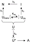
Proposed folding mechanism for bhx showing a parallel refolding pathway which results from proline isomerization, as well as formation of an off-pathway oligomer. Parentheses indicate folding steps which occur primarily in the unfolded state.

References

    1. Dobson CM, Protein misfolding, evolution and disease, Trends Biochem. Sci 24 (1999) 329–332. - PubMed
    1. Wetzel R, Ideas of order for amyloid fibril structure, Structure 10 (2002) 1031–1036. - PubMed
    1. Govaerts C, Wille H, Prusiner SB, Cohen FE, Evidence for assembly of prions with left-handed {beta}-helices into trimers, Proc. Nat. Acad. Sci. U. S. A 101 (2004) 8342–8347. - PMC - PubMed
    1. Weigele PR, Scanlon E, King J, Homotrimeric, beta-stranded viral adhesins and tail proteins, J. Bacteriol 185 (2003) 4022–4030. - PMC - PubMed
    1. Junker M, Schuster CC, McDonnell AV, Sorg KA, Finn MC, Berger B, Clark PL, Pertactin beta-helix folding mechanism suggests common themes for the secretion and folding of autotransporter proteins, Proc. Natl. Acad. Sci. U. S. A 103 (2006) 4918–4923. - PMC - PubMed

Publication types

LinkOut - more resources