. 2009 Apr 24;284(17):11267-78.
doi: 10.1074/jbc.M807211200. Epub 2009 Mar 2.

Unfractionated heparin and new heparin analogues from ascidians (chordate-tunicate) ameliorate colitis in rats

Affiliations

Unfractionated heparin and new heparin analogues from ascidians (chordate-tunicate) ameliorate colitis in rats

Celso L R Belmiro et al. J Biol Chem. .

Abstract

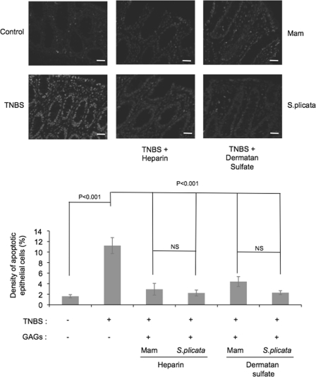
The anti-inflammatory effect of mammalian heparin analogues, named dermatan sulfate and heparin, isolated from the ascidian Styela plicata was accessed in a TNBS-induced colitis model in rats. Subcutaneous administration of the invertebrate compounds during a 7-day period drastically reduced inflammation as observed by the normalization of the macroscopic and histological characteristics of the colon. At the molecular level, a decrease in the production of TNF-alpha, TGF-beta, and VEGF was observed, as well as a reduction of NF-kappaB and MAPK kinase activation. At the cellular level, the heparin analogues attenuated lymphocyte and macrophage recruitment and epithelial cell apoptosis. A drastic reduction in collagen-mediated fibrosis was also observed. No hemorrhagic events were observed after glycan treatment. These results strongly indicate the potential therapeutic use of these compounds for the treatment of colonic inflammation with a lower risk of hemorrhage when compared with mammalian heparin.


Figures

FIGURE 1.
FIGURE 1.
Effect of heparin analogues on histological parameters of inflamed colon. Colonic samples from Wistar rats after TNBS-induced colitis without or with heparin or ascidian heparin analogues administration were fixed in 40 g/liter formaldehyde saline and stained with HE (A). Unfractionated mammalian heparin (porcine intestinal mucosa) or ascidian heparin analogues (dermatan sulfate and heparin from S. plicata) (8 mg/kg/day) were administered to the animals subcutaneously for 7 days. The colonic samples were scored according to the following histological parameters: ulceration, hyperplasia, and inflammatory infiltrate. For both inflammatory infiltrate and hyperplasia, grading was considered: 3, severe; 2, moderate; 1, mild; 0, absent. For ulcers, grading was: 4, diffuse glandular disruption or extensive deep ulceration; 3, glandular disruption or focal deep ulceration; 2, diffuse superficial ulceration; 1, focal superficial ulceration; and 0, absent (B). In control experiments, the histological evaluation was performed after treatment of the ascidian GAGs with nitrous acid (HONO) or chondroitin ABC lyase (Chase ABC) (C). Values are mean ± S.E. of 10 animals/group. Statistical differences among the experimental groups were evaluated with the one-way ANOVA test. The level of significance was set at p < 0.05. Mam, mammalian. Magnification, ×100. Scale bar, 20 μm.
FIGURE 2.
FIGURE 2.
Effect of heparin analogues on macrophage infiltration into the inflamed colon. Colonic samples from Wistar rats after TNBS-induced colitis without or with heparin or ascidian heparin analogues administration were immediately embedded in Tissue-Tek O.C.T. compound, snap-frozen in isopentane in a liquid nitrogen bath, and submitted to immunohistochemical analysis using mouse monoclonal anti-rat ED1. Heparin and heparin analogues were administered to the animals as described in the legend of Fig. 1. The number of immunoreactive cells per millimeter-squared was counted in at least 10 different areas. Quantitative analysis of tissue sections were carried out under light microscopy at ×400 magnification. Values are mean ± S.E. of 10 animals/group. Statistical differences among the experimental groups were evaluated with the one-way ANOVA test. The level of significance was set at p < 0.05. Mam, mammalian. Scale bar, 20 μm.
FIGURE 3.
FIGURE 3.
Effect of heparin analogues on lymphocyte infiltration into the inflamed colon. Colonic samples from Wistar rats after TNBS-induced colitis without or with heparin or ascidian heparin analogues administration were immediately embedded in Tissue-Tek O.C.T. compound, snap-frozen in isopentane in a liquid nitrogen bath, and submitted to immunohistochemical analysis using mouse monoclonal anti-rat CD3. Heparin and heparin analogues were administered to the animals as described in the legend of Fig. 1. The number of immunoreactive cells per millimeter-squared was counted in at least 10 different areas. Quantitative analysis of tissue sections were carried out under light microscopy at ×400 magnification. Values are mean ± S.E. of 10 animals/group. Statistical differences among the experimental groups were evaluated with the one-way ANOVA test. The level of significance was set at p < 0.05. Mam, mammalian. Scale bar, 20 μm.
FIGURE 4.
FIGURE 4.
Effect of heparin analogues on cytokine production in the inflamed colon. Colonic mucosal explants from Wistar rats after TNBS-induced colitis without or with heparin or ascidian heparin analogues administration were cultured for 24 h at 37 °C. After centrifugation, the supernatants were used for measurement of the concentration of cytokines by a commercial sensitive enzyme-linked immunosorbent assay (ELISA) method for rat TNF-α, TGF-β, and VEGF, as described under “Experimental Procedures.” Heparin and heparin analogues were administered to the animals as described in the legend of Fig. 1. Quantitative analysis of tissue sections were carried out under light microscopy at ×400 magnification. Values were expressed as picogram of cytokine/mg protein and represent the mean ± S.E. of 10 animals/group. Statistical differences among the experimental groups were evaluated with the one-way ANOVA test. The level of significance was set at p < 0.05. Mam, mammalian. Scale bar, 20 μm.
FIGURE 5.
FIGURE 5.
Effect of heparin analogues on NF-κB activation in the inflamed colon. Colonic samples from Wistar rats after TNBS-induced colitis without or with heparin or ascidian heparin analogues administration were immediately embedded in Tissue-Tek O.C.T. compound, snap-frozen, and submitted to immunohistochemical analysis using mouse monoclonal anti-rat p65. Heparin and heparin analogues were administered to the animals as described in the legend of Fig. 1. Quantitative analysis of tissue sections were carried out under light microscopy at ×800 magnification. The number of cells with nuclear NF-κB-staining (NF-κB-positive cells) per millimeter-squared was counted in at least 10 different areas. Values are mean ± S.E. of 10 animals/group. Statistical differences among the experimental groups were evaluated with the one-way ANOVA test. The level of significance was set at p < 0.05. Mam, mammalian. Scale bar, 20 μm.
FIGURE 6.
FIGURE 6.
Effect of heparin analogues on ERK activation in the inflamed colon. Colonic samples from Wistar rats after TNBS-induced colitis without or with heparin or ascidian heparin analogues administration were immediately embedded in Tissue-Tek O.C.T. compound, snap-frozen in isopentane in a liquid nitrogen bath, and submitted to immunohistochemical analysis using mouse monoclonal anti-rat P-ERK1/2 and FITC-conjugated goat anti-mouse IgG antibody. Heparin and heparin analogues were administered to the animals as described in the legend of Fig. 1. The tissues were observed with a Zeiss LSM 510 confocal laser scanning microscope. The number of immunoreactive cells in the lamina propria per millimeter-squared was counted in at least 10 different areas. Values are mean ± S.E. of 10 animals/group. Statistical differences among the experimental groups were evaluated with the one-way ANOVA test. The level of significance was set at p < 0.05. Mam, mammalian. Scale bar, 20 μm.
FIGURE 7.
FIGURE 7.
Effect of heparin analogues on collagen deposition in the inflamed colon. Colonic samples from Wistar rats after TNBS-induced colitis without or with heparin or ascidian heparin analogues administration were fixed in 40 g/liter formaldehyde saline and the collagen fibers stained with phosphomolibidic acid-picro-sirius red dye. Heparin and heparin analogues were administered to the animals as described in the legend of Fig. 1. Density of collagen fibers was defined by the area positively stained for collagen in relation to total intestinal tissue per millimeter-squared using an imaging analysis system. At least 15 different areas per tissue section were analyzed under light microscopy at ×400 magnification. Values are mean ± S.E. The level of significance was set at p < 0.05. Mam, mammalian. Scale bar, 20 μm.
FIGURE 8.
FIGURE 8.
Effect of heparin analogues on epithelial cell apoptosis in the inflamed colon. Colonic samples from Wistar rats after TNBS-induced colitis without or with heparin or ascidian heparin analogues administration were embedded in paraffin and apoptotic cells were determined by the TUNEL assay, as described under “Experimental Procedures.” Heparin and heparin analogues were administered to the animals as described in the legend of Fig. 1 and sections were analyzed in a confocal microscope. The density of apoptotic cells was defined as the percentage of immunoreactive cells within at least 500 epithelial cells in the crypts and in the surface epithelium of longitudinally sectioned colonic pits. Values are mean ± S.E. The level of significance was set at p < 0.05. Mam, mammalian. Magnification ×400. Scale bar, 20 μm.
FIGURE 9.
FIGURE 9.
Effect of ascidian heparin on LPS-induced macrophage activation. Rat peritoneal macrophages were obtained as described under “Experimental Procedures.” Cells were incubated in 24-well tissue culture plates for 24 h in the presence of a range of heparin concentrations (5.0, 10, 20, and 40 μg/ml) plus LPS (1 μg/ml) (Sigma), added 2 h after heparin treatment (A). In some experiments LPS was omitted to check the effect of heparin on the macrophages (B). The concentration of TNF-α was determined by enzyme-linked immunosorbent assay kit (R&D Systems). The data shown are the average of two independent experiments. Each point was performed in duplicate.
FIGURE 10.
FIGURE 10.
Effect of heparin analogues on coagulation and platelet counts ex vivo. At experimental day 7, animals from the different groups were anesthetized and blood samples were collected. A, aPTT was carried out in rat plasma using a coagulometer (Amelung KC4A) and expressed in seconds, as described under “Experimental Procedures.” B, platelet count in the whole blood was measured on an automatic hematology analyzer. Results are expressed as number of cells per cubic millimeter. Values are mean ± S.E. The level of significance was set at p < 0.05. Mam, mammalian.

Similar articles

Cited by

References

    1. Xavier, R. J., and Podolsky, D. K. (2007) Nature 448, 427-434 - PubMed
    1. Papadakis, K., and Targan, S. (2000) Inflamm. Bowel Dis. 6, 303-313 - PubMed
    1. Reinecker, H., Steffen, M., Witthoeft, T., Pflueger, I., Schreiber, S., MacDermott, R., and Raedler, A. (1993) Clin. Exp. Immunol. 94, 174-181 - PMC - PubMed
    1. Fiocchi, C. (1998) Gastroenterology 115, 182-205 - PubMed
    1. Podolsky, D. (2002) N. Engl. J. Med. 347, 417-429 - PubMed

Publication types