. 2009 May 1;284(18):12178-87.
doi: 10.1074/jbc.M900994200. Epub 2009 Mar 3.

Identification of GRO1 as a critical determinant for mutant p53 gain of function

Affiliations

Identification of GRO1 as a critical determinant for mutant p53 gain of function

Wensheng Yan et al. J Biol Chem. .

Abstract

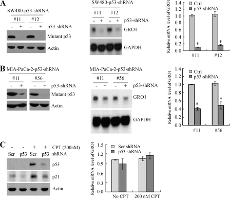
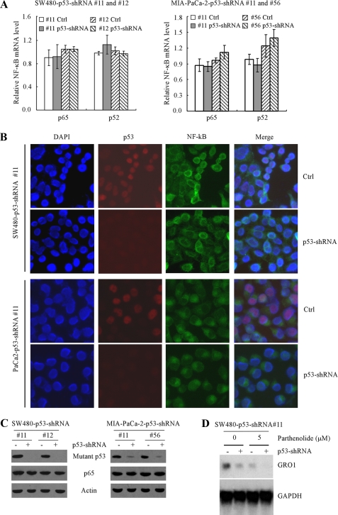
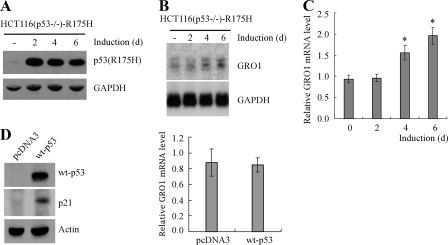
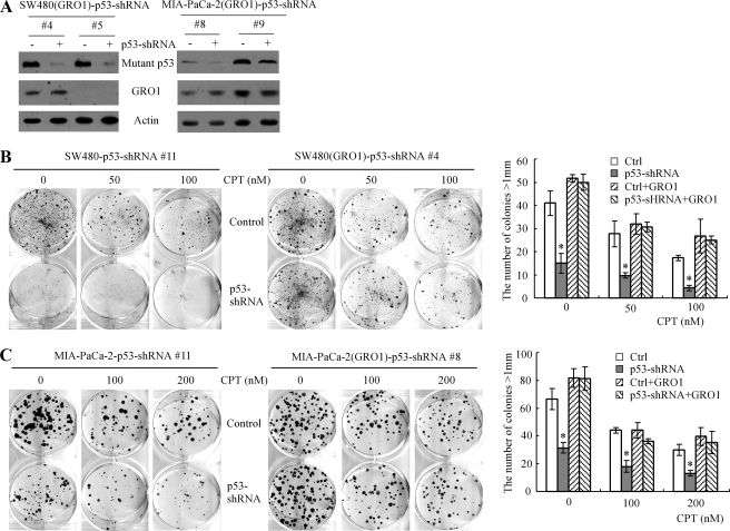
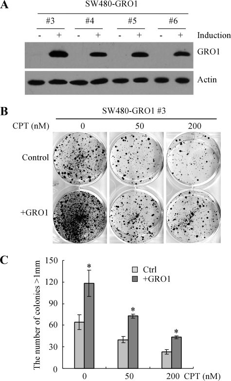
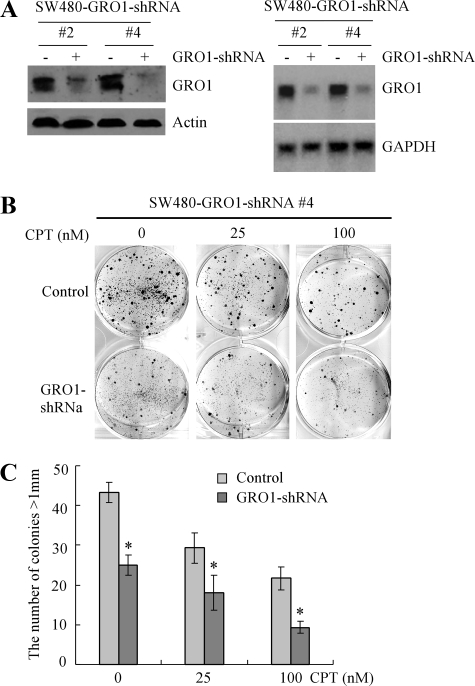
Mutant p53 gain of function contributes to cancer progression, increased invasion and metastasis potentials, and resistance to anticancer therapy. The ability of mutant p53 to acquire its gain of function is shown to correlate with increased expression of progrowth genes, such as c-MYC, MDR1, and NF-kappaB2. However, most of the published studies to identify mutant p53 target genes were performed in a cell system that artificially overexpresses mutant p53. Thus, it remains unclear whether such mutant p53 targets can be regulated by endogenous physiological levels of mutant p53. Here, we utilized SW480 and MIA-PaCa-2 cells, in which endogenous mutant p53 can be inducibly knocked down, to identify mutant p53 target genes that potentially mediate mutant p53 gain of function. We found that knockdown of mutant p53 inhibits GRO1 expression, whereas ectopic expression of mutant R175H in p53-null HCT116 cells increases GRO1 expression. In addition, we found that endogenous mutant p53 is capable of binding to and activating the GRO1 promoter. Interestingly, ectopic expression of GRO1 can rescue the proliferative defect in SW480 and MIA-PaCa-2 cells induced by knockdown of mutant p53. Conversely, knockdown of endogenous GRO1 inhibits cell proliferation and thus abrogates mutant p53 gain of function in SW480 cells. Taken together, our findings define a novel mechanism by which mutant p53 acquires its gain of function via transactivating the GRO1 gene in cancer cells. Thus, targeting GRO1 for cancer therapy would be applicable to a large portion of human tumors with mutant p53, but the exploration of GRO1 as a potential target should take the mutation status of p53 into consideration.


Figures

FIGURE 1.
FIGURE 1.
Knockdown of mutant p53 inhibits GRO1 expression in SW480 and MIA-PaCa-2 cells. A, left, generation of SW480 cell lines in which mutant p53 can be inducibly knocked down. Western blots were prepared with extracts from SW480 cells uninduced (-) or induced (+) to knock down mutant p53 for 3 days and then probed with antibodies against p53 and actin, respectively. Middle, knockdown of mutant p53 in SW480 cells down-regulates GRO1 expression. Northern blots were prepared with total RNAs isolated from SW480 cells treated as above. The blots were probed with cDNAs derived from the GRO1 and GAPDH genes, respectively. The level of GAPDH mRNA was measured as a loading control. Right, the relative levels of GRO1 transcript were measured by quantitative reverse transcription-PCR and normalized by levels of GAPDH mRNA. B, left, generation of MIA-PaCa-2 cell lines in which mutant p53 can be inducibly knocked down. Western blot analyses were performed as in A. Middle and right, knockdown of mutant p53 in MIA-PaCa-2 cells down-regulates GRO1 expression. Northern blot and quantitative reverse transcription-PCR analyses were performed as in A. C, left, transient knockdown of wild-type p53 has no effect on Gro1 expression in HCT116 cells. Western blots were prepared with extracts from HCT116 cells with transient knockdown of wild-type p53 for 3 days and then probed with antibodies against p53, p21, and actin, respectively. A scrambled shRNA was used as a negative control. Right, the relative levels of GRO1 transcript in HCT116 cells with or without wild-type p53 knockdown were quantified by real-time PCR.
FIGURE 2.
FIGURE 2.
Down-regulation of GRO1 induced by mutant p53 knockdown is independent of the NF-κB pathway in SW480 and MIA-PaCa-2 cells. A, knockdown of mutant p53 had no obvious effect on the levels of p65 and p52 mRNA in SW480 cells (left) and MIA-PaCa-2 cells (right). Quantitative reverse transcription-PCR was performed with total RNAs isolated from SW480 and MIA-PaCa-2 cells that were uninduced or induced to knock down mutant p53 for 3 days. B, knockdown of mutant p53 had no obvious effect on the localization of p65 protein in SW480 cells (top two panels) and MIA-PaCa-2 cells (bottom two panels). Immunofluorescence staining was performed to detect the cellular location of p65 and p53 as described under “Experimental Procedures.” C, knockdown of mutant p53 had no obvious effect on the levels of p65 protein in SW480 cells (left) and MIA-PaCa-2 cells (right). Western blots were prepared with extracts from SW480 and MIA-PaCa-2 cells treated as in A and then probed with antibodies against p53, p65, and actin, respectively. D, knockdown of mutant p53 decreased the level of GRO1 mRNA regardless of inhibition of the NF-κB pathway. SW480-p53-shRNA cells (clone 11) were uninduced (-) or induced (+) to knock down mutant p53 along with treatment of 0 or 5 μm parthenolide, a specific inhibitor of the NF-κB pathway. The levels of GRO1 mRNA were measured by Northern blot analysis as in Fig. 1.
FIGURE 3.
FIGURE 3.
GRO1 expression was increased by ectopic expression of mutant p53 (R175H) in p53-null HCT116 cells. A, Western blots were prepared with extracts from p53-null HCT116 cells uninduced (-) or induced to express mutant R175H for 2, 4, or 6 days and then probed with antibodies against p53 and actin, respectively. B, endogenous GRO1 expression was increased by ectopic expression of mutant R175H. Northern blots were prepared with total RNAs purified from p53-null HCT116 cells uninduced (-) or induced to express mutant p53 for 2, 4, or 6 days and probed with cDNAs derived from the GRO1 and GAPDH genes, respectively. C, the relative levels of GRO1 transcript in p53-null HCT116 cells induced to express R175H for 0–6 days were quantified by real-time reverse transcription-PCR. D, left, Western blots were prepared with extracts from p53-null HCT116 cells with or without transient overexpression of wild-type p53 for 4 days. Right, the relative levels of GRO1 transcript in p53-null HCT116 cells in the presence or absence of wild-type p53 for 4 days were quantified by real-time PCR.
FIGURE 4.
FIGURE 4.
Mutant p53 can bind to and transactivate the GRO1 promoter. A, schematic presentation of the GRO1 (top), p21 (middle), and GAPDH (bottom) promoters and the location of PCR primers used for ChIP assay. B, the binding of mutant p53 to the GRO1 promoter in SW480 cells was measured by ChIP. Mutant p53-DNA complexes were captured with anti-p53 antibody along with rabbit IgG as a control. The binding of mutant p53 protein to the p21 and GAPDH promoters was also measured as negative controls. C, pull-down assay of mutant p53 (top) and NF-κB (p65) (bottom) binding to the GRO1 promoter in vitro. The biotinated GRO1 promoter probe bound to streptavidin-agarose beads was used to precipitate p65 and mutant p53 from nuclear protein extracts from SW480 cells uninduced (-) or induced (+) to knock down mutant p53 for 3 days. The biotinated GAPDH promoter probe was used as a negative binding control. The nuclear proteins pulled down by biotinated promoter probes were analyzed by Western blot assay with antibodies against p53 and p65, respectively. D, schematic presentation of the luciferase reporter constructs under the control of the GRO1 (top) and p21 (bottom) promoters, respectively. E, the GRO1 promoter is responsive to mutant p53-R273H (left) but not wild-type p53 (right). The response of the p21 promoter to wild-type p53 was measured as a positive control. The luciferase activity for the promoterless pGL2 plasmid in the presence of wild-type and mutant p53 was measured as a negative control. Luciferase assay was performed as described under “Experimental Procedures.”
FIGURE 5.
FIGURE 5.
Ectopic expression of GRO1 can rescue the proliferative defect induced by mutant p53 knockdown. A, generation of SW480 and MIA-PaCa-2 cell lines in which GRO1 was stably expressed and endogenous mutant p53 can be inducibly knocked down. Western blots were prepared with extracts from SW480 and MIA-PaCa-2 cells uninduced (-) or induced (+) to knock down mutant p53 for 3 days and then probed with antibodies against p53 and actin, respectively. B and C, left, knockdown of mutant p53 inhibits colony formation in SW480 cells (B) and MIA-PaCa2 cells (C) that were mock-treated or treated with 50–200 nm camptothecin (CPT). Middle, ectopic expression of GRO1 rescues the proliferative defect induced by mutant p53 knockdown in SW480 cells (B) and MIA-PaCa2 cells (C) that were mock-treated or treated with 50–200 nm camptothecin. Right, quantification of the number of colonies with a diameter of >1 mm. *, p < 0.05.
FIGURE 6.
FIGURE 6.
Ectopic expression of GRO1 promotes cell proliferation in SW480 cells. A, generation of SW480 cell lines in which GRO1 can be inducibly expressed under the control of the teteracycline-regulated promoter. Western blots were prepared with extracts from SW480 cells uninduced (-) or induced (+) to express GRO1 for 24 h and then probed with antibodies against GRO1 and actin, respectively. B, ectopic expression of GRO1 alone is capable of promoting cell proliferation in SW480 cells. SW480-GRO1 cells were uninduced or induced to express GRO1 along with treatment of 0, 50, or 200 nm CPT, cultured for ∼3 weeks, and then fixed and stained to measure the number and size of colonies formed. C, quantification of the number of colonies with a diameter of >1 mm shown in B.*, p < 0.05.
FIGURE 7.
FIGURE 7.
Knockdown of endogenous GRO1 inhibits cell proliferation in SW480 cells. A, generation of SW480 cell lines in which endogenous GRO1 can be inducibly knocked down. Left, Western blots were prepared with extracts from SW480 cells uninduced (-) and induced (+) to knock down GRO1 for 3 days and then probed with antibodies against GRO1 and actin, respectively. Right, Northern blots were prepared with total RNAs purified from SW480 cells uninduced (-) and induced (+) to knock down GRO1 for 3 days and probed with cDNAs derived from the GRO1 and GAPDH genes, respectively. B, knockdown of GRO1 inhibits colony formation in SW480 cells. SW480 cells were uninduced or induced to knock down GRO1 along with treatment of 0, 25, or 100 nm CPT, cultured for ∼3 weeks, and then fixed and stained to measure the number and size of colonies formed. C, quantification of the number of colonies with a diameter of >1 mm shown in B.*, p < 0.05.

Similar articles

Cited by

References

    1. Kalo, E., Buganim, Y., Shapira, K. E., Besserglick, H., Goldfinger, N., Weisz, L., Stambolsky, P., Henis, Y. I., and Rotter, V. (2007) Mol. Cell. Biol. 27 8228-8242 - PMC - PubMed
    1. Bossi, G., Lapi, E., Strano, S., Rinaldo, C., Blandino, G., and Sacchi, A. (2006) Oncogene 25 304-309 - PubMed
    1. Atema, A., and Chene, P. (2002) Cancer Lett. 185 103-109 - PubMed
    1. Frazier, M. W., He, X., Wang, J., Gu, Z., Cleveland, J. L., and Zambetti, G. P. (1998) Mol. Cell. Biol. 18 3735-3743 - PMC - PubMed
    1. Lane, B. R., Liu, J., Bock, P. J., Schols, D., Coffey, M. J., Strieter, R. M., Polverini, P. J., and Markovitz, D. M. (2002) J. Virol. 76 11570-11583 - PMC - PubMed

Publication types