. 2009 May 8;284(19):12762-71.
doi: 10.1074/jbc.M806084200. Epub 2009 Mar 3.

NLRP3/cryopyrin is necessary for interleukin-1beta (IL-1beta) release in response to hyaluronan, an endogenous trigger of inflammation in response to injury

Affiliations

NLRP3/cryopyrin is necessary for interleukin-1beta (IL-1beta) release in response to hyaluronan, an endogenous trigger of inflammation in response to injury

Kenshi Yamasaki et al. J Biol Chem. .

Abstract

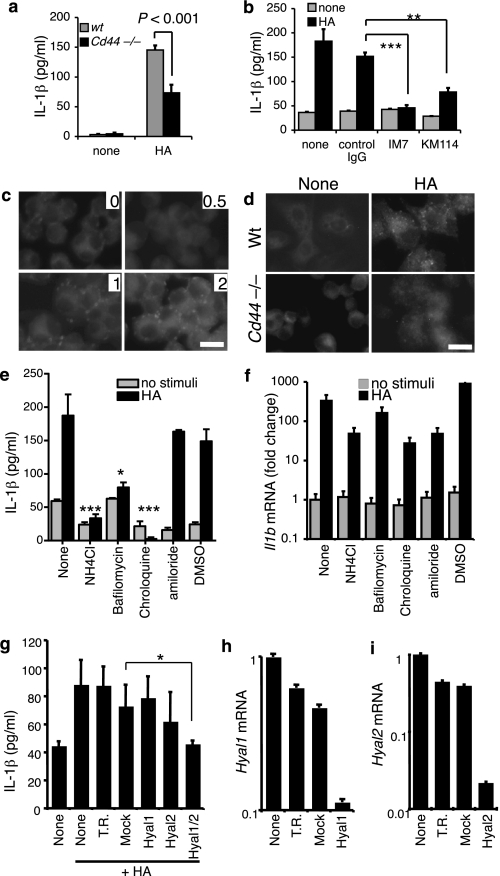
Inflammation under sterile conditions is a key event in autoimmunity and following trauma. Hyaluronan, a glycosaminoglycan released from the extracellular matrix after injury, acts as an endogenous signal of trauma and can trigger chemokine release in injured tissue. Here, we investigated whether NLRP3/cryopyrin, a component of the inflammasome, participates in the inflammatory response to injury or the cytokine response to hyaluronan. Mice with a targeted deletion in cryopyrin showed a normal increase in Cxcl2 in response to sterile injuries but had decreased inflammation and release of interleukin-1beta (IL-1beta). Similarly, the addition of hyaluronan to macrophages derived from cryopyrin-deficient mice increased release of Cxcl2 but did not increase IL-1beta release. To define the mechanism of hyaluronan-mediated activation of cryopyrin, elements of the hyaluronan recognition process were studied in detail. IL-1beta release was inhibited in peritoneal macrophages derived from CD44-deficient mice, in an MH-S macrophage cell line treated with antibodies to CD44, or by inhibitors of lysosome function. The requirement for CD44 binding and hyaluronan internalization could be bypassed by intracellular administration of hyaluronan oligosaccharides (10-18-mer) in lipopolysaccharide-primed macrophages. Therefore, the action of CD44 and subsequent hyaluronan catabolism trigger the intracellular cryopyrin --> IL-1beta pathway. These findings support the hypothesis that hyaluronan works through IL-1beta and the cryopyrin system to signal sterile inflammation.


Figures

FIGURE 1.
FIGURE 1.
Sterile injury induced less IL-1β in cryopyrin null mouse. a, cryopyrin null (Nlrp3–/–) and WT mice were injured by liquid nitrogen or sterile bead injection as described under “Experimental Procedures,” and skin erythema was monitored. Representative images of the skin surface 48 h after injury from three independents experiments are shown. b and c, injured skin of cryopyrin null (ko) and WT mouse was excised, and IL-1β (b) and Cxcl2/MIP-2 (c) were measured by ELISA. Bars indicate the mean of each group. No-treat, non-treated. **: p < 0.01. d, TLR4 null (ko) and WT mice were injured by liquid nitrogen or sterile bead injection. Skin lesions were excised, and IL-1β was measured by ELISA. Bars indicate the mean of each group. *: p < 0.05, **: p < 0.01.
FIGURE 2.
FIGURE 2.
IL-1β release by HA depends on cryopyrin. a and b, peritoneal macrophages from cryopyrin null (Nlrp3–/–) and WT control mice were treated with HA (25 μg/ml) or LPS (25 μg/ml) for 18 h, and IL-1β (a) and Cxcl2/MIP-2 (b) in cultured media were measured by ELISA. Mean and S.E. are shown. gray bars, WT; black bars, cryopyrin null (Nlrp3–/–). Statistical analyses were done to compare Nlrp3–/– and WT in each stimulus. ***: p < 0.001. c–f, peritoneal macrophages from cryopyrin null (Nlrp3–/–) and WT control mice were treated with various concentrations of HA for 18 h. IL-1β (c) and Cxcl2/MIP-2 (d) release in cultured media was measured by ELISA. mRNA abundance for Il1b (e) and Cxcl2 (f) was measured 18 h after exposure to various doses of HA by quantitative RT-PCR. Quantitative RT-PCR data are shown as relative expression as compared with control untreated WT peritoneal macrophages. Statistical analyses were done to compare Nlrp3–/– and WT in each concentration. *: p < 0.05, ***: p < 0.001. g and h, peritoneal macrophages from TLR4 null (Tlr4–/–) and WT mice were treated with HA (25 μg/ml) for 18 h, and IL-1β in cultured media (g) and the level of Il1b mRNA (h) were measured. mRNA was measured by quantitative RT-PCR and shown as relative expression over the control (Wt indicates non-treated). Mean and S.E. are shown on the graphs. **: p < 0.01, ***: p < 0.001.
FIGURE 3.
FIGURE 3.
CD44 affects IL-1β release in response to HA. a, peritoneal macrophages from CD44-deficient mice and WT mice were treated with HA (25 μg/ml) for 18 h, and IL-1β in cultured media was measured by ELISA. Mean ± S.E. are shown. b, MH-S macrophage cell line was pretreated with rat anti-mouse CD44 monoclonal antibodies KM114 and IM7 or control rat IgG (10 μg/ml each) for 30 min. MH-S cells were then exposed to 25 μg/ml HA for 18 h in the presence of antibodies. IL-1β in cultured media was measured by ELISA. **: p < 0.01, ***: p < 0.001. c, MH-S cells were treated with 1 μm of LysoSensor (Green DND-189, Invitrogen) for 30 min and washed once with PBS. After the addition of HA (25 μg/ml), fluorescence was monitored at the time points indicated by the digits in the upper right corner that denote hours after HA stimulation. Acidic intracellular compartments, which are indicated by green fluorescence, were observed at 1 and 2 h after HA addition. Scale bar, 50 μm. d, peritoneal macrophages from CD44-deficient mice and WT mice were treated with 1 μm of LysoSensor for 30 min and washed once with PBS. Two hours after the addition of HA (25 μg/ml), fluorescence were monitored. Scale bar, 50 μm. e and f, MH-S cells were pretreated with the indicated agents or DMSO (0.1%) as vehicle control for 30 min. Then MH-S cells were treated with 25 μg/ml HA for 18 h in the presence of agents, and IL-1β in cultured media (e) and Il1b mRNA (f) was measured. Mean and S.E. are shown. The agents used were: lysosome inhibitors NH4Cl (20 mm), bafilomycin A1 (100 nm), chroloquine (100 μm), and Na+–H+ exchanger inhibitor amiloride (500 μm). Statistical analyses were done to compare each vehicle-treated sample. *: p < 0.05, **: p < 0.01, ***: p < 0.001. g, MH-S cells were treated with siRNAs for 72 h. Then the cells were treated with 25 μg/ml HA for 18 h in the presence of agents, and IL-1β in cultured media was measured. Mean and S.E. are shown. The agents used were: none, no pretreatment; T.R., transfection reagent alone; Mock, mock transfected; Hyal1, Hyal1 siRNA-transfected; Hyal2, Hyal2 siRNA-transfected; Hyal1/2, both Hyal1 and Hyal2 siRNA-transfected. *: p < 0.05. h and i, MH-S cells were treated with siRNAs for 72 h, and Hyal1 (h) and Hyal2 (i) mRNA expression was examined.
FIGURE 4.
FIGURE 4.
CD44 is involved in HA endocytosis. a, MH-S cells were treated with fl-HA (25 μg/ml) for 24 h. Scale bar, 50 μm. b, peritoneal macrophages from WT and Cd44–/– mice were treated with fl-HA for the indicated times. Numbers indicate the percentage of fluorescein-positive cells (percentage of fl-HA-positive cells/4′,6-diamidino-2-phenylindole (DAPI)). Scale bar, 50 μm.
FIGURE 5.
FIGURE 5.
Intracellular HA oligosaccharides (HA oligo) induced IL-1β release from LPS-primed macrophages. a–d, MH-S cells were stimulated with HA oligosaccharides (100 μm) or HA (25 μg/ml) for 18 h, and IL-1β (a) and Cxcl2/MIP-2 (b) in cultured media were measured. mRNA abundance for Il1b (c) and Cxcl2 (d) at 18 h was measured by quantitative RT-PCR. Quantitative RT-PCR data are shown as relative expression as compared with untreated control (none). Mean and S.E. are shown on the graphs. Statistical analyses were done to compare with non-treated samples. **: p < 0.01. e–h, MH-S cells were pretreated with or without LPS (0.2 μg/ml) for 3 h. Then media were replaced, and 2.5 μg of HA oligosaccharides were added to the media or permitted access to the intracellular space by membrane transfection with DOTAP. IL-1β (e) and Cxcl2/MIP-2 (f) in cultured media were measured after 18 h stimulation. mRNA abundance for Il1b (c) and Cxcl2 (d) at 18 h are shown as relative expression as compared with untreated control (none). Mean and S.E. are shown on the graphs. Statistical analyses were done to compare each vehicle-treated sample. *: p < 0.05, **: p < 0.01, ***: p < 0.001.
FIGURE 6.
FIGURE 6.
A model of HA catabolism and inflammasome activation. HA binding to CD44 activates p38 mitogen-activated protein kinase (MAPK) and NF-κB through TLR4 to increase IL-1β and Cxcl2 mRNA transcription. Endocytosis of CD44 and hyaluronidase activity hydrolyzes HA to small oligosaccharide fragments. Small HA oligosaccharides activate the inflammasome through NLRP3/cryopyrin to convert pro-IL-1β to active IL-1β and release IL-1β.

Similar articles

Cited by

References

    1. Beutler, B. (2004) Nature 430 257–263 - PubMed
    1. Beg, A. A. (2002) Trends Immunol. 23 509–512 - PubMed
    1. Johnson, G. B., Brunn, G. J., Kodaira, Y., and Platt, J. L. (2002) J. Immunol. 168 5233–5239 - PubMed
    1. Termeer, C., Benedix, F., Sleeman, J., Fieber, C., Voith, U., Ahrens, T., Miyake, K., Freudenberg, M., Galanos, C., and Simon, J. C. (2002) J. Exp. Med. 195 99–111 - PMC - PubMed
    1. Tammi, R., Saamanen, A. M., Maibach, H. I., and Tammi, M. (1991) J. Investig. Dermatol. 97 126–130 - PubMed

Publication types

MeSH terms