. 2009 Apr 1;122(Pt 7):905-11.
doi: 10.1242/jcs.028183. Epub 2009 Mar 3.

Cell polarity triggered by cell-cell adhesion via E-cadherin

Affiliations

Cell polarity triggered by cell-cell adhesion via E-cadherin

Ravi A Desai et al. J Cell Sci. .

Abstract

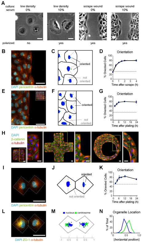
Cell polarity is orchestrated by numerous extracellular cues, and guides events such as chemotaxis, mitosis and wound healing. In scrape-wound assays of cell monolayers, wound-edge cells orient their centrosomes towards the wound, a process that appears to depend on the formation of new cell-extracellular-matrix adhesions as cells spread into the wound. In direct contrast to scrape-wounded cells, isolated cells without cell-cell contacts failed to polarize, suggesting that asymmetry of cell-cell adhesions resulting from monolayer disruption might contribute to polarization. By using micropatterned substrates to engineer such asymmetries in kidney epithelial cells, we found that cell-cell contact induced displacement of the nucleus towards the contact, and also caused centrosomal reorientation and lamellipodial ruffling to the distal side of the nucleus. Upon release from micropatterned constraints, cells exhibited directed migration away from the cell-cell contact. Disrupting E-cadherin engagement randomized nuclear position and lamellipodial ruffling in patterned cultures, and abrogated scrape-wound-induced cell reorientation, but not migration rate. Polarity that was induced by cell-cell contact required an intact actin cytoskeleton and Cdc42 activity, but not RhoA or Rac signaling. Together, these findings demonstrate a novel role for cell-cell adhesion in polarization, and have implications for wound healing and developmental patterning.


Figures

Fig. 1.
Fig. 1.
Cell-cell contact polarizes the cell-motility apparatus. (A) Cells seeded in 10% serum at subconfluence or confluence were switched to 0% or 10% serum-containing media and imaged 33 hours later (5 hours after the scrape wound). (B) Cells 4 hours after scrape wounding. (C) Edge cells were scored as being oriented if their centrosome was distal from cell-cell contacts relative to the nucleus. (D) Kinetics of orientation. Broken line indicates random (33%) orientation; t=0 corresponds to time of scrape wounding. (E) Cells in a patterned monolayer 24 hours after plating. Inset is a 250,000 μm2 patterned monolayer and shows the region of the main panel. (F) Edge orientation as in C. (G) Kinetics of orientation as in D, except that t=0 corresponds to cell plating. (H) Cells on patterns 24 hours after plating. Boxed areas are shown at higher magnification on the right of each panel. (I) Cells in a paired configuration 24 hours after plating. (J) Orientation as in C. (K) Kinetics of orientation as in D, except that t=0 corresponds to cell plating and the broken line indicates random (50%) orientation. (L) Cells in a paired configuration 8 hours after plating. (M) Pattern (gray), nuclear (blue) and centrosomal (green) positions were measured 12 hours after plating from micrographs of 50 patterns from three experiments. (N) Frequency histogram of organelle locations. In D, G and K, means ± s.e.m. are from at least three independent experiments. Scale bars: 25 μm.
Fig. 2.
Fig. 2.
Cell-cell contact polarizes cell dynamics. (A) Representative image and kymographs of cell membranes that are distal (i) or proximal (ii) to the cell-cell contact from a 15-minute time-series. In the kymographs, vertical and horizontal bars are 10 μm and 60 seconds, respectively. (B, left) Activity map of the cell pair in A. (B, right) Dynamic activity per pixel of membrane that is proximal (dashed boxes in the left image) or distal (solid box in the left image) to the cell-cell contact for 20 cells. Means (horizontal bars) ± s.e.m. (gray boxes) are indicated (*P=0.0112; Student's t-test). (C) Representative time series of a pair of cells after release (time indicated) from patterned constraints (broken lines) 20 hours after plating. Scale bars: 25 μm unless noted.
Fig. 3.
Fig. 3.
E-cadherin is required for cell-cell-contact-induced polarity. (A) Cells were switched to minimum essential medium modified for suspension cultures (SMEM) containing 1.8 or 0 mM Ca2+ at 4 hours, and fixed at 24 hours, after plating; processed as in Fig. 1I-K (*P=0.018; Student's t-test). (B,C) Cells infected with Ad-GFP or Ad-EΔ at least 6 hours prior to, and fixed 24 after, plating; processed as in Fig. 1I-N (**P=0.0032; Student's t-test). (D) Representative images and kymographs from a 15-minute time series. In the kymographs, vertical and horizontal bars are 10 μm and 60 seconds, respectively. (E) Activity maps of the cell pairs in D, and dynamic activity per pixel of membrane that is proximal (dotted boxes) or distal (solid box) to the cell-cell contact for 20 cells (Ad-GFP, green) and nine cells (Ad-EΔ, blue). Means ± s.e.m. are indicated (*P<0.05; ns, P>0.05; ANOVA and Tukey's HSD). (F,G) Cells were infected at least 24 hours prior to wounding and imaged subsequently (F) or immunolabeled and scored 4 hours after wounding (G) (ns, P=0.45; **P=0.0031; Student's t-test). The broken line indicates random orientation, and means ± s.e.m. are from at least three independent experiments. Scale bars: 25 μm unless noted.
Fig. 4.
Fig. 4.
Polarity triggered by cell-cell contact requires the actin cytoskeleton and Cdc42. (A-D) G0-synchronized cells were treated with 1% DMSO or 1 μM cytochalasin D (cyto. D) at 3 hours, and fixed at 8 hours, after plating; processed as in Fig. 1I-N (**P=0.0072; Student's t-test). (E) Cells were infected with Ad-GFP or Ad-RhoN19 at least 12 hours prior to, and fixed 24 hours after, plating; processed as in Fig. 1I-K (ns, P=1; Student's t-test). (F) Cells were handled as in A, except that they were treated with PBS or 50 μM NSC23766 (ns, P=0.17; Student's t-test). (G) Representative immunoblot and quantification of total and GTP-bound Cdc42, measured via pull-down assay from cells transfected with 10 nM siRNA (*P<0.05; ns, P>0.05; ANOVA and Tukey's HSD). (H-J) siRNA-transfected cells fixed 8 hours after plating and processed as in Fig. 1I-N (*P<0.05; ns, P>0.05; ANOVA and Tukey's HSD). Broken lines in C, E, F and I indicate random orientation, and means ± s.e.m. are from at least three independent experiments. Scale bars: 25 μm.

References

    1. Adams, C. L., Chen, Y. T., Smith, S. J. and Nelson, W. J. (1998). Mechanisms of epithelial cell-cell adhesion and cell compaction revealed by high-resolution tracking of E-cadherin-green fluorescent protein. J. Cell Biol. 142, 1105-1119. - PMC - PubMed
    1. Braga, V. M. and Yap, A. S. (2005). The challenges of abundance: epithelial junctions and small GTPase signalling. Curr. Opin. Cell Biol. 17, 466-474. - PubMed
    1. Chu, Y. S., Thomas, W. A., Eder, O., Pincet, F., Perez, E., Thiery, J. P. and Dufour, S. (2004). Force measurements in E-cadherin-mediated cell doublets reveal rapid adhesion strengthened by actin cytoskeleton remodeling through Rac and Cdc42. J. Cell Biol. 167, 1183-1194. - PMC - PubMed
    1. Drees, F., Pokutta, S., Yamada, S., Nelson, W. J. and Weis, W. I. (2005). Alpha-catenin is a molecular switch that binds E-cadherin-beta-catenin and regulates actin-filament assembly. Cell 123, 903-915. - PMC - PubMed
    1. Ehrlich, J. S., Hansen, M. D. and Nelson, W. J. (2002). Spatio-temporal regulation of Rac1 localization and lamellipodia dynamics during epithelial cell-cell adhesion. Dev. Cell 3, 259-270. - PMC - PubMed

Publication types