. 2009 Mar 15;69(6):2615-22.
doi: 10.1158/0008-5472.CAN-08-3022. Epub 2009 Mar 3.

NKX3.1 activates expression of insulin-like growth factor binding protein-3 to mediate insulin-like growth factor-I signaling and cell proliferation

Affiliations

NKX3.1 activates expression of insulin-like growth factor binding protein-3 to mediate insulin-like growth factor-I signaling and cell proliferation

Erin Muhlbradt et al. Cancer Res. .

Abstract

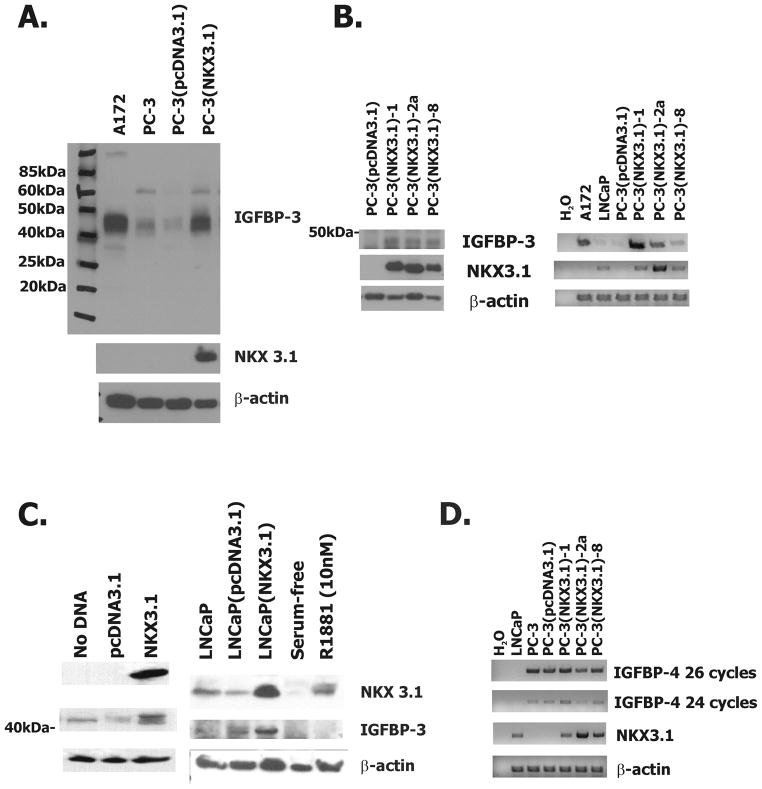
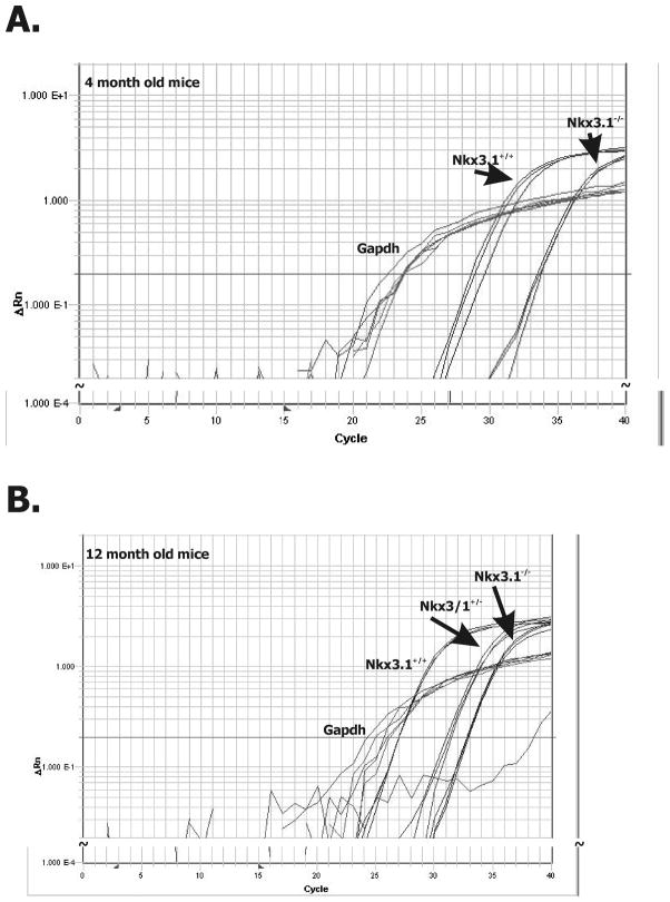
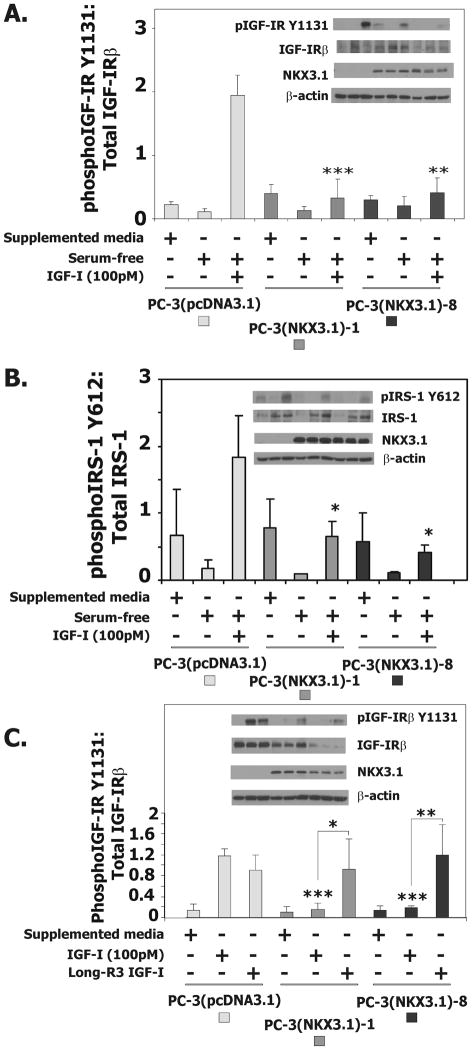
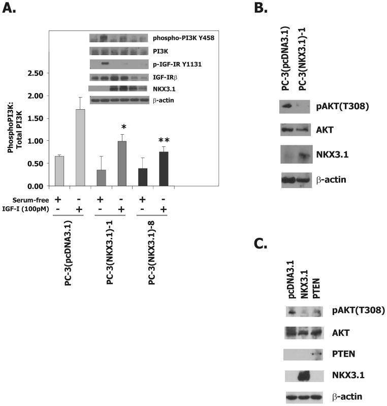
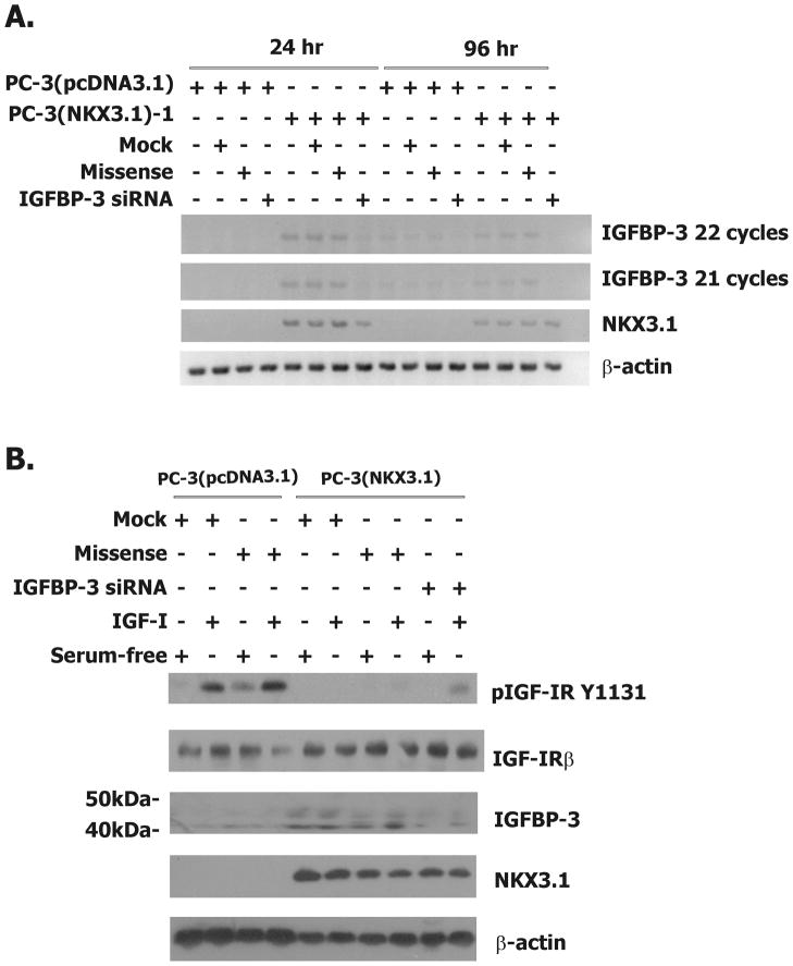
NKX3.1 is a homeobox gene that codes for a haploinsufficient prostate cancer tumor suppressor. NKX3.1 protein levels are down-regulated in the majority of primary prostate cancer tissues. NKX3.1 expression in PC-3 cells increased insulin-like growth factor binding protein-3 (IGFBP-3) mRNA expression 10-fold as determined by expression microarray analysis. In both stably and transiently transfected PC-3 cells and in LNCaP cells, NKX3.1 expression increased IGFBP-3 mRNA and protein expression. In prostates of Nkx3.1 gene-targeted mice Igfbp-3 mRNA levels correlated with Nkx3.1 copy number. NKX3.1 expression in PC-3 cells attenuated the ability of insulin-like growth factor-I (IGF-I) to induce phosphorylation of type I IGF receptor (IGF-IR), insulin receptor substrate 1, phosphatidylinositol 3-kinase, and AKT. The effect of NKX3.1 on IGF-I signaling was not seen when cells were exposed to long-R3-IGF-I, an IGF-I variant peptide that does not bind to IGFBP-3. Additionally, small interfering RNA-induced knockdown of IGFBP-3 expression partially reversed the attenuation of IGF-IR signaling by NKX3.1 and abrogated NKX3.1 suppression of PC-3 cell proliferation. Thus, there is a close relationship in vitro and in vivo between NKX3.1 and IGFBP-3. The growth-suppressive effects of NKX3.1 in prostate cells are mediated, in part, by activation of IGFBP-3 expression.


Figures

Figure 1
Figure 1
NKX3.1 up regulates the expression of IGFBP-3 in prostate cancer cell lines. A, Western blot analysis of cell extracts from stably expressing PC-3(pcDNA3.1) and PC-3(NKX3.1)-1 cell clones that have been analyzed by expression array. B, On the left is western blot analysis of extracts from additional PC-3 clones expressing NKX3.1. One the right is RT-PCR analysis of PC-3(pcDNA3.1) and PC-3(NKX3.1) clones for NKX3.1 and IGFBP-3 expression. A172 cell extract is a positive control for IGFBP-3 expression and LNCaP cell extract is used as a positive control for NKX3.1 expression. C, On the left is western blot analysis of extracts from PC-3 cells transiently transfected with an NKX3.1 expression vector. On the right is western blot analysis of extracts from LNCaP cells transiently transfected with an NKX3.1 expression vector or serum starved in 5% CCS supplemented media overnight and treated with 10nM R1881. D, RT-PCR analysis of IGFBP-4 mRNA expression in PC-3(pcDNA3.1) and PC-3(NKX3.1) clones.
Figure 2
Figure 2
NKX3.1 and IGFBP-3 expression in mouse prostate. Real-time RT-PCR analysis of mRNA extracts of prostatic tissue of Nkx3.1 gene targeted mice at (A) 4 and (B) 12 months of age. Anterior prostates were analyzed from three separate animals of each genotype at each time point. Gapdh expression was used as a loading control. Each one of the multiple assay lines in each group represent the results of a single tissue sample. The results correlated with Nkx3.1 genotype.
Figure 3
Figure 3
NKX3.1 expression inhibits the IGF-1-mediated phosphorylation of the IGF-1R and IRS-1 in PC-3 cells. A, Western blot analysis of extracts from PC-3(pcDNA3.1), PC-3(NKX3.1)-1 and PC-3(NKX3.1)-8 stable cell clones serum starved for 16 hours and treated with 100pM IGF-I. The histogram of IGF-IR activation is based upon three separate experiments. B, Western blot analysis of extracts from PC-3(pcDNA3.1), PC-3(NKX3.1)-1 and PC-3(NKX3.1)-8 clones serum starved for 16 hours and treated with 100pM IGF-I. The histogram of IRS-1 activation is based upon three separate experiments. C, Western blot analysis of extracts from PC-3(pcDNA3.1), PC-3(NKX3.1)-1, and PC-3(NKX3.1)-8 clones serum starved for 16 hours and treated with 100pM IGF-1 or Long-R3-IGF-I. The histogram of IGF-IR activation is based upon three separate experiments. Statistical comparisons are indicated by asterisks as explained in Materials and Methods. Comparisons are versus PC-3(pcDNA3.1) treated with IGF-I unless otherwise indicate by brackets.
Figure 4
Figure 4
NKX3.1 attenuates IGF-1R downstream signaling. A, Western blot analysis of extracts from PC-3(pcDNA3.1), PC-3(NKX3.1)-1, and PC-3(NKX3.1)-8 clones serum starved for 16 hours and treated with 100pM IGF-I for 3 minutes. The histogram of PI-3K activation is based upon three separate experiments. Statistical comparisons are indicated by asterisks. Comparisons are versus PC-3(pcDNA3.1) treated with IGF-I. B, Western blot analysis of cell extracts from PC-3(pcDNA3.1) and PC-3(NKX3.1)-1 cells grown in media containing 10% FBS. C, Western blot analysis of cell extracts of PC-3 cells transiently transfected with either a NKX3.1 or a PTEN expression vector.
Figure 5
Figure 5
IGFBP-3 knock down in PC-3(NKX3.1) cells. A, RT-PCR analysis of mRNA from PC-3(pcDNA3.1) and PC-3(NKX3.1)-1 cells that were treated with transfection reagent alone, missense oligo, or the IGFBP-3 siRNA oligo at 24 and 96 hours post-transfection. B. Western blot analysis of extracts from PC-3(pcDNA3.1) and PC-3(NKX3.1)-1 cells treated with transfection reagent alone, missense oligo, or the IGFBP-3 siRNA oligo for 96 hours, after which they were serum starved for 16 hours and treated with 100pM IGF-I.

Similar articles

Cited by

References

    1. Swalwell JI, Vocke CD, Yang Y, et al. Determination of a minimal deletion interval on chromosome band 8p21 in sporadic prostate cancer. Genes Chromosomes Cancer. 2002;33:201–5. - PubMed
    1. Bowen C, Bubendorf L, Voeller HJ, et al. Loss of NKX3. 1 expression in human prostate cancers correlates with tumor progression. Cancer Res. 2000;60:6111–5. - PubMed
    1. Asatiani E, Huang WX, Wang A, et al. Deletion, methylation, and expression of the NKX3. 1 suppressor gene in primary human prostate cancer. Cancer Res. 2005;65:1164–73. - PubMed
    1. Voeller HJ, Augustus M, Madlike V, et al. Coding region of NKX3. 1, prostate-specific homeobox gene on 8p21, is not mutated in human prostate cancers. Cancer Res. 1997;57:4455–9. - PubMed
    1. Ornstein DK, Cinquanta M, Weiler S, et al. Expression studies and mutational analysis of the androgen regulated homeobox gene nkx3. 1 in benign and malignant prostate epithelium. J Urol. 2001;165:1329–34. - PubMed

Publication types

MeSH terms

Substances