Review
. 2009 Mar;73(1):36-61.
doi: 10.1128/MMBR.00026-08.

Biochemical features and functional implications of the RNA-based T-box regulatory mechanism

Affiliations
Review

Biochemical features and functional implications of the RNA-based T-box regulatory mechanism

Ana Gutiérrez-Preciado et al. Microbiol Mol Biol Rev. 2009 Mar.

Abstract

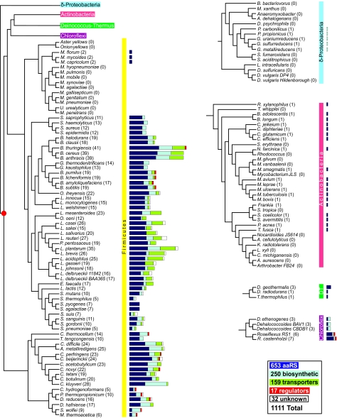
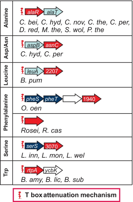
The T-box mechanism is a common regulatory strategy used for modulating the expression of genes of amino acid metabolism-related operons in gram-positive bacteria, especially members of the Firmicutes. T-box regulation is usually based on a transcription attenuation mechanism in which an interaction between a specific uncharged tRNA and the 5' region of the transcript stabilizes an antiterminator structure in preference to a terminator structure, thereby preventing transcription termination. Although single T-box regulatory elements are common, double or triple T-box arrangements are also observed, expanding the regulatory range of these elements. In the present study, we predict the functional implications of T-box regulation in genes encoding aminoacyl-tRNA synthetases, proteins of amino acid biosynthetic pathways, transporters, and regulatory proteins. We also consider the global impact of the use of this regulatory mechanism on cell physiology. Novel biochemical relationships between regulated genes and their corresponding metabolic pathways were revealed. Some of the genes identified, such as the quorum-sensing gene luxS, in members of the Lactobacillaceae were not previously predicted to be regulated by the T-box mechanism. Our analyses also predict an imbalance in tRNA sensing during the regulation of operons containing multiple aminoacyl-tRNA synthetase genes or biosynthetic genes involved in pathways common to more than one amino acid. Based on the distribution of T-box regulatory elements, we propose that this regulatory mechanism originated in a common ancestor of members of the Firmicutes, Chloroflexi, Deinococcus-Thermus group, and Actinobacteria and was transferred into the Deltaproteobacteria by horizontal gene transfer.


Figures

FIG. 1.
FIG. 1.
The T-box RNA regulatory system. (A) Structural model of the B. subtilis tyrS T-box leader RNA. The T-box element present in the B. subtilis tyrS leader region was originally described by Grundy and Henkin (see reference 43). The standard T-box leader RNA arrangement consists of three major elements, stem I, stem II, and stem III plus the stem IIA/stem IIB pseudoknot, and the competing terminator and antiterminator structures. The specifier loop, an internal bulge in stem I, contains the specifier sequence (boxed UAC residues complementary to the anticodon sequence of tRNATyr); the conserved purine (an adenine) following the specifier sequence is inside a green circle. The T-box sequence is unpaired in the terminator form and is paired in the antiterminator form (the antiterminator is shown to the right of the terminator). The sequence highlighted in blue shows the nucleotides involved in the antiterminator structure. The antiterminator structure has a bulge that interacts with the unpaired residues at the acceptor end of an uncharged tRNA. Nucleotide conservation in all 722 T-box sequences analyzed was evaluated using a multiple sequence alignment obtained from the Rfam database (42), and residues are color coded accordingly. (B) Model of the regulatory alternatives for the T-box mechanism. During the transcription of a leader region by RNA polymerase (red ovals), the nascent RNA folds into a structure competent for binding of the cognate tRNA at two sites. The binding of uncharged tRNA (top) to both the specifier sequence and the antiterminator bulge stabilizes the antiterminator (green RNA segment), preventing the formation of the terminator. This allows transcription to proceed into the downstream-regulated coding sequence (blue box). Charged tRNA (represented by Tyr attached to the 3′ end of the tRNA) can interact with the specifier sequence but cannot interact with the antiterminator; a failure to stabilize the antiterminator allows the formation of the terminator helix (red RNA segment), and transcription is terminated before the downstream coding region can be transcribed. Conserved elements of T-box RNAs are stem I (black), stem II (orange), the stem IIA/stem IIB pseudoknot (light blue), and stem III (purple).
FIG. 2.
FIG. 2.
Distribution of T-box regions in different phylogenetic taxa. The phylogenetic tree for organisms relevant to our study was constructed based on the phylogenetic distances of aligned sequences from the concatenation of 31 proteins in 191 species, as previously described (18). Alignments were generated using the program MUSCLE (28), and phylogenetic reconstruction was performed using the PROTDIST program of the PHYLIP phylogeny inference package program (version 3.57c; J. Felsenstein, University of Washington, Seattle). Operons were predicted based on an analysis of intergenic distances, as described previously (76). Horizontal bar lengths are drawn to scale, reflecting the number of operons regulated by a T-box sequence; these are classified into one of the following groups: aminoacyl-tRNA synthetases (dark blue), amino acid biosynthetic genes (light blue), genes coding for regulatory proteins (red), transporter genes (green), and genes of unknown function (white). The most parsimonious scenario would place the initially evolved T-box regulatory sequence in a common ancestor of the Firmicutes, the Actinobacteria, the Chloroflexi, and the Deinococcus-Thermus (DT) group. The postulated origin is represented by a red dot in the tree. Names of Firmicutes are as follows: M. hyopneumoniae, Mycoplasma hyopneumoniae; M. pulmonis, Mycoplasma pulmonis; M. mobile, Mycoplasma mobile; M. synoviae, Mycoplasma synoviae; M. agalactiae, Mycoplasma agalactiae; M. gallisepticum, Mycoplasma gallisepticum; M. genitalium, Mycoplasma genitalium; M. pneumoniae, Mycoplasma pneumoniae; U. urealyticum, Ureaplasma urealyticum; M. penetrans, Mycoplasma penetrans. Names of Deltaproteobacteria are as follows: B. bacteriovorus, Bdellovibrio bacteriovorus; M. xanthus, Myxococcus xanthus; A. dehalogenans, Anaeromyxobacter dehalogenans; D. psychrophila, Desulfotalea psychrophila; S. fumaroxidans, Syntrophobacter fumaroxidans; S. aciditrophicus, Syntrophus aciditrophicus; L. intracellularis, Lawsonia intracellularis; D. sulfuricans, Desulfovibrio desulfuricans; D. vulgaris, Desulfovibrio vulgaris. Names of Actinobacteria are as follows: T. whipplei, Tropheryma whipplei; M. gilvum, Mycobacterium gilvum; M. vanbaalenii, Mycobacterium vanbaalenii; S. tropica, Salinispora tropica; A. cellulolyticus, Acidothermus cellulolyticus; K. radiotolerans, Kineococcus radiotolerans; L. xyli, Leifsonia xyli; C. michiganensis, Clavibacter michiganensis; A. aurescens, Arthrobacter aurescens. Additional names are listed in the legend to Fig. 3.
FIG. 3.
FIG. 3.
Aminoacyl-tRNA synthetase genes regulated by the T-box mechanism. From the set of T-box-regulated genes identified in our study, operons containing aaRS genes were grouped according to the amino acid class of the aaRS. Operons containing more than one different aaRS gene are shown under the amino acid category matching the predicted specifier sequence. In the exceptional case of leuS in C. hydrogenoformans, P. thermopropionicum, and S. wolfei, the T-box sequence, drawn in green, contains a tRNA gene. Organism nomenclature is as follows for members of the Firmicutes: A. met, “Alkaliphilus metalliredigens”; B. amy, Bacillus amyloliquefaciens; B. ant, Bacillus anthracis; B. cla, Bacillus clausii; B. cer, Bacillus cereus; B. hal, Bacillus halodurans; B. lic, Bacillus lichenformis; B. pum, Bacillus pumilus; B. sub, Bacillus subtilis; B. ste, Bacillus stearothermophilus; B. thu, Bacillus thuringiensis; C. ace, Clostridium acetobutylicum; C. bei, Clostridium beijerinckii; C. bot, Clostridium botulinum; C. dif, Clostridium difficile; C. hyd, Clostridium hydrogenoformans; C. klu, Clostridium kluyveri; C. nov, Clostridium novyi; C. per, Clostridium perfringens; C. tet, Clostridium tetani; C. the, Clostridium thermocellum; D. haf, Desulfitobacterium hafniense; D. red, Desulfotomaculus reducens; E. fae, Enterococcus faecalis; G. kau, Geobacillus kaustophilus; G. the, Geobacillus thermodenitrificans; L. aci, Lactobacillus acidophilus; L. bre, Lactobacillus brevis; L. cas, Lactobacillus casei; L. del, both Lactobacillus delbrueckii subsp. bulgaricus strains; L. del 11842, Lactobacillus delbrueckii subsp. bulgaricus strain ATCC 11842; L. del BAA365, Lactobacillus delbrueckii subsp. bulgaricus strain ATCC BAA365; L. gas, Lactobacillus gasseri; L. inn, Listeria innocua; L. joh, Lactobacillus johnsonii; L. lac, Lactococcus lactis; L. mes, Leuconostoc mesenteroides; L. mon, Listeria monocytogenes; L. pla, Lactobacillus plantarum; L. reu, Lactobacillus reuteri; L. sak, Lactobacillus sakei; L. sal, Lactobacillus salivarius; L. wel, Listeria welshimeri; M. cap, Mycoplasma capricolum; M. flo, Mesoplasma florum; M. myc, Mycoplasma mycoides; M. the, Moorella thermoacetica; O. ihe, Oceanobacillus iheyensis; O. oen, Oenococcus oeni; P. pen, Pediococcus pentosaceus; P. the, Pelotomaculum thermopropionicum; S. aga, Streptococcus agalactiae; S. aur, Staphylococcus aureus; S. epi, Staphylococcus epidermidis; S. gor, Streptococcus gordonii; S. hae, Staphylococcus haemolyticus; S. mut, Streptococcus mutans; S. pne, Streptococcus pneumoniae; S. pyo, Streptococcus pyogenes; S. san, Streptococcus sanguinis; S. sap, Staphylococcus saprophyticus; S. sui, Streptococcus suis; S. the, Streptococcus thermophilus; S. wol, Syntrophomonas wolfei; T. ten, Thermoanaerobacter tengcongensis. Organism nomenclature is as follows for members of the Actinobacteria: B. ado, Bifidobacterium adolescentis; B. lon, Bifidobacterium longum; C. eff, Corynebacterium efficiens; C. dip, Corynebacterium diphtheriae; C. glu, Corynebacterium glutamicum; C. jei, Corynebacterium jeikeium; M. avi, Mycobacterium avium; M. bov, Mycobacterium bovis; M. lep, Mycobacterium leprae; M. sme, Mycobacterium smegmatis; M. tub, Mycobacterium tuberculosis; M. ulc, Mycobacterium ulcerans; N. far, Nocardia farcinica; P. can, Propionibacterium acnes; R. xyl, Rubrobacter xylanophilus; S. ave, Streptomyces avermitilis; S. coe, Streptomyces coelicolor; T fus, Thermobifida fusca. Organism nomenclature is as follows for members of the Fusobacteria: F. nuc, Fusobacterium nucleatum. Organism nomenclature is as follows for members of the Deinococcus-Thermus group: D. geo, Deinococcus geothermalis; D. rad, Deinococcus radiodurans; T. the, Thermus thermophilus. Organism nomenclature is as follows for members of the Chlorobi: C. tep, Chlorobium tepidum; C. aur, Chloroflexus aurantiacus; C. hut, Cytophaga hutchinsonii. Organism nomenclature is as follows for members of the Chloroflexi: D. BAV1, “Dehalococcoides” sp. strain BAV1; D. CBDB1, Dehalococcoides sp. strain CBDB1; D. eth, “Dehalococcoides ethenogenes”; R. SM1, Roseiflexus sp. strain SM1; R. cas, Roseiflexus castenholzii. Organism nomenclature is as follows for members of the Proteobacteria: G. sul, Geobacter sulfurreducens; G. met, Geobacter metallireducens; G. ura, Geobacter uraniumreducens; P. car, Pelobacter carbinolicus; P. pro, Pelobacter propionicus. Operon predictions and the color code used for the different types of regulated genes are described in the legend of Fig. 2.
FIG. 4.
FIG. 4.
Variety of mechanisms used in regulating methionine and serine biosynthetic genes of B. subtilis and other bacteria. The regulatory mechanisms and operon arrangements found in B. subtilis (left column) are compared with those of T-box-regulated operons in other members of the Firmicutes (right column). Where genes of other organisms share the same regulatory mechanism as B. subtilis, the names of these organisms are indicated in the B. subtilis column. The graphic representation of each type of regulatory element is indicated in the red box in each subfigure. No attempt was made to identify pathways exhibiting feedback inhibition of enzyme activity; only those reported in the literature for B. subtilis are indicated. The regulatory proteins shown represent their corresponding binding sites in the operon. Genes that have not been annotated were labeled based on their corresponding COG numbers (i.e., a gene that belongs to COG1878 is drawn as an arrow containing the number “1878”). Organism abbreviations and color codes are described in the legends of Fig. 2 and 3.
FIG. 5.
FIG. 5.
Variety of mechanisms used in regulating leucine, isoleucine, valine, and histidine biosynthetic genes of B. subtilis and other bacteria. The color code used for the different types of regulated genes and abbreviations of organisms are described in the legend of Fig. 2. The graphic representation of each type of regulatory element is described in the legend of Fig. 4.
FIG. 6.
FIG. 6.
Variety of mechanisms used in regulating alanine, arginine, asparagine, aspartate, cysteine, glycine, and threonine biosynthetic genes of B. subtilis and other bacteria. The color code used for the different types of regulated genes and abbreviations of organisms are described in the legend of Fig. 2. The graphic representation of each type of regulatory element is described in the legend of Fig. 4.
FIG. 7.
FIG. 7.
Variety of mechanisms used in regulating phenylalanine, proline, tryptophan, and tyrosine biosynthetic genes of B. subtilis and other bacteria. The color code used for the different types of regulated gene and abbreviations of organisms are described in the legend of Fig. 2. The graphic representation of each type of regulatory element is described in the legend of Fig. 4. TRAP, in addition to transcriptionally regulating the trp operon in B. subtilis and its closest relatives, can also regulate trpE translation by binding to the trpE leader RNA and promoting the formation of a secondary structure that sequesters the SD sequence, inhibiting translation initiation (73).
FIG. 8.
FIG. 8.
Genes involved in amino acid transport that are regulated by the T-box mechanism. The common designation for each class of transporter gene is shown inside each arrow. Genes that have not been annotated were labeled based on their corresponding COG numbers (i.e., a gene that belongs to COG4166 is drawn as an arrow with the number “4166”). Note that genes are named in accordance with the GenBank annotation and might not represent the real specificity of the transporter as revealed by the identification of the specifier codon in our T-box analysis. Organism abbreviations and gene color codes are described in the legends of Fig. 2 and 3.
FIG. 9.
FIG. 9.
Common strategy in regulating amino acid transporter genes and biosynthetic genes. Both amino acid transporters and biosynthetic enzymes can fulfill the need of an organism for certain amino acids. This results in a tendency to coordinate the expression of the genes for these classes of proteins by using a shared regulatory mechanism. As shown for three amino acid-related genes, this tendency is specific for each phylogenetic clade. The regulatory elements were identified using our Riboswitch Web server (RibEx) (1) and previously reported data (2). [1], the SMK riboswitch regulates metK genes in lactic acid bacteria, including Enterococcus, Streptococcus, and Lactococcus spp. (see reference 33). [2], in streptococci, unlike other members of the Firmicutes, methionine biosynthesis and transport are controlled by protein transcription factors, in this case, MtaR, MetR, and CmbR (see reference 67). Organism names and gene color codes are described in the legends of Fig. 2 and 3.
FIG. 10.
FIG. 10.
Regulatory genes controlled by the T-box mechanism. Organism abbreviations and color codes are described in the legends of Fig. 2 and 3. COG1940 is annotated as a “negative regulator of the xylose operon”; this annotation does not correspond to the function deduced from its Phe T-box specificity. COG3070 corresponds to a “regulator of competence-specific genes”; its true function is unknown, but it is predicted to be related to its Ser specifier sequence and the fact that is cotranscribed with serS. COG2207 is the family of AraC transcriptional regulators. The Roseiflexus (Rosei) sp. regulatory genes do not belong to any COG family but are annotated in GenBank as “putative transcriptional regulators, MerR family.”

Similar articles

Cited by

References

    1. Abreu-Goodger, C., and E. Merino. 2005. RibEx: a Web server for locating riboswitches and other conserved bacterial regulatory elements. Nucleic Acids Res. 33W690-W692. - PMC - PubMed
    1. Abreu-Goodger, C., N. Ontiveros-Palacios, R. Ciria, and E. Merino. 2004. Conserved regulatory motifs in bacteria: riboswitches and beyond. Trends Genet. 20475-479. - PubMed
    1. Ahn, K. S., and R. G. Wake. 1991. Variations and coding features of the sequence spanning the replication terminus of Bacillus subtilis 168 and W23 chromosomes. Gene 98107-112. - PubMed
    1. Alifano, P., R. Fani, P. Lio, A. Lazcano, M. Bazzicalupo, M. S. Carlomagno, and C. B. Bruni. 1996. Histidine biosynthetic pathway and genes: structure, regulation, and evolution. Microbiol. Rev. 6044-69. - PMC - PubMed
    1. Babitzke, P., and P. Gollnick. 2001. Posttranscription initiation control of tryptophan metabolism in Bacillus subtilis by the trp RNA-binding attenuation protein (TRAP), anti-TRAP, and RNA structure. J. Bacteriol. 1835795-5802. - PMC - PubMed

Publication types

MeSH terms

LinkOut - more resources