Review
. 2009 Mar;73(1):155-77.
doi: 10.1128/MMBR.00024-08.

Pivotal roles of the outer membrane polysaccharide export and polysaccharide copolymerase protein families in export of extracellular polysaccharides in gram-negative bacteria

Affiliations
Review

Pivotal roles of the outer membrane polysaccharide export and polysaccharide copolymerase protein families in export of extracellular polysaccharides in gram-negative bacteria

Leslie Cuthbertson et al. Microbiol Mol Biol Rev. 2009 Mar.

Abstract

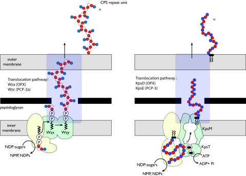
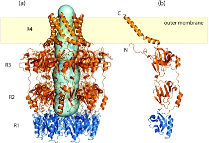
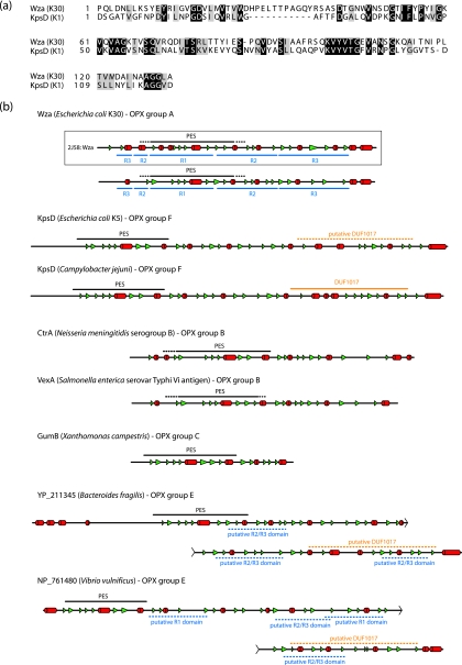
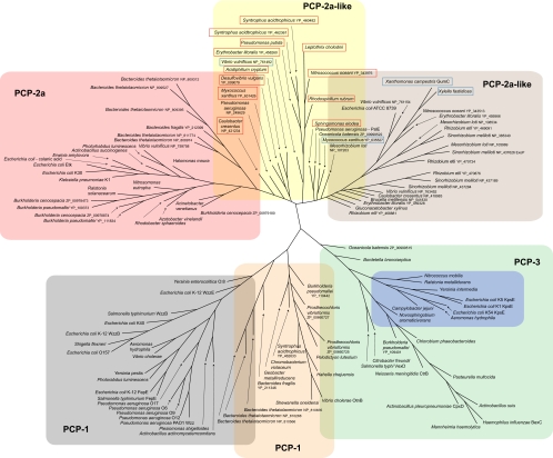
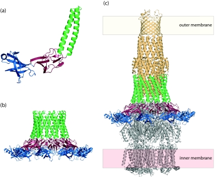
Many bacteria export extracellular polysaccharides (EPS) and capsular polysaccharides (CPS). These polymers exhibit remarkably diverse structures and play important roles in the biology of free-living, commensal, and pathogenic bacteria. EPS and CPS production represents a major challenge because these high-molecular-weight hydrophilic polymers must be assembled and exported in a process spanning the envelope, without compromising the essential barrier properties of the envelope. Emerging evidence points to the existence of molecular scaffolds that perform these critical polymer-trafficking functions. Two major pathways with different polymer biosynthesis strategies are involved in the assembly of most EPS/CPS: the Wzy-dependent and ATP-binding cassette (ABC) transporter-dependent pathways. They converge in an outer membrane export step mediated by a member of the outer membrane auxiliary (OMA) protein family. OMA proteins form outer membrane efflux channels for the polymers, and here we propose the revised name outer membrane polysaccharide export (OPX) proteins. Proteins in the polysaccharide copolymerase (PCP) family have been implicated in several aspects of polymer biogenesis, but there is unequivocal evidence for some systems that PCP and OPX proteins interact to form a trans-envelope scaffold for polymer export. Understanding of the precise functions of the OPX and PCP proteins has been advanced by recent findings from biochemistry and structural biology approaches and by parallel studies of other macromolecular trafficking events. Phylogenetic analyses reported here also contribute important new insight into the distribution, structural relationships, and function of the OPX and PCP proteins. This review is intended as an update on progress in this important area of microbial cell biology.


Figures

FIG. 1.
FIG. 1.
Morphology of CPS- and EPS-producing bacteria. (a) K. pneumoniae serotype K20; (b) E. coli serotype K30. The EPS and CPS in these bacteria have identical repeat-unit structures, and both forms are labeled using cationized ferritin. The E. coli isolate retains most of the polymer as CPS in a well-defined capsule structure. In contrast, the K. pneumoniae isolate has some CPS but releases substantial amounts of polymer from the surface as EPS; this “looser” association is evident in the micrograph.
FIG. 2.
FIG. 2.
Overview of the Wzy-dependent (left) and ABC transporter-dependent (right) EPS biosynthesis pathways. The two biosynthesis pathways involve different components, modes of polymerization, and membrane topologies (for details, see the text). However, both involve terminal export pathways mediated by members of the PCP and OPX protein families.
FIG. 3.
FIG. 3.
Structure of Wza from E. coli K30, the prototype OPX protein. (a) Wza forms an octameric complex composed of four ring domains (R1 to R4), with the R4 domain spanning the outer membrane. The internal cavity of the Wza complex was calculated using the online Caver tool (http://loschmidt.chemi.muni.cz/caver/) and is indicated as a pale blue surface. (b) A single Wza monomer (Protein Data Bank accession no. 2J58). The R1 domain (dark blue) contains the majority of the OPX-specific PES motif and likely represents the minimal structural unit of the PES motif. Structure diagrams were generated using Pymol (DeLano Scientific LLC).
FIG. 4.
FIG. 4.
Predicted secondary structures of OPX proteins indicate a significant degree of structural similarity between the various homologs. (a) Alignment of the primary sequences of the PES domains from Wza and E. coli KpsD (an OPX protein from the ABC transporter-dependent group 2 CPS system). (b) Predicted secondary structures from Wza and other OPX representatives. The secondary structures were assigned based on the agreement of at least three out of four structure prediction algorithms (Prof [82], PSIPred [13, 44], SSPro [16], and jPRED [18]). α-Helical structures are indicated as red cylinders, and β-structures are depicted by green arrowheads. The OPX-specific PES motifs (as detected by a conserved-domain search [65]) are shown as a black line above the secondary structural elements, and the dashed sections of these lines represent regions where no significant sequence alignment to the canonical motif could be found. The DUF1017 motif in the KpsD protein of C. jejuni was identified in a motif search. The “putative DUF1017” domain (hatched orange lines) in the E. coli KpsD protein was assigned based on similar predicted secondary structure. Several regions in the “long” OPX from B. fragilis (YP_211345) showed sequence similarity to the R2 and R3 domains from Wza. These regions were designated “putative R2/R3 domains” (hatched blue lines) if their predicted secondary structure matched the corresponding domains in the Wza prototype. The secondary structure for the solved crystal structure of Wza (bounded by a box) has been included as a measure of the accuracy of the predicted secondary structure assignments. In all cases, only the mature proteins are shown; signal sequences have been removed.
FIG. 5.
FIG. 5.
Phylogenetic tree showing the relationship between OPX proteins. Sequences were identified using key word and BLAST searches (2). Only the PES motif of each OPX was used in the analysis. Alignments were performed using CLUSTALW (57). Phylogenetic analysis was carried out on 100 bootstrapped data sets using the parsimony program in the PHYLIP package (30). Trees were viewed using SplitsTree (41). The tree is based on data available in September 2008.
FIG. 6.
FIG. 6.
The predicted secondary structures of the periplasmic regions of PCP proteins demonstrate the high degree of α-helical content found in the different PCP families. The structures were calculated using the approach used for the OPX proteins in Fig. 4. The transmembrane regions were assigned based on the agreement of three transmembrane prediction algorithms (TMHMM [105], TMPred [39], and TMPro [33]). The secondary structures from solved crystal structures of the periplasmic domains of the Wzz homologs, WzzST from S. enterica serovar Typhimurium (Protein Data Bank accession no. 3B8P) and WzzFepE from E. coli O157:H7 (Protein Data Bank accession no. 3B8M), are bounded by black boxes.
FIG. 7.
FIG. 7.
Structures of PCP-1 proteins. The figure shows data obtained for FepE (Protein Data Bank accession no. 3B8M) from E. coli O157:H7 (112). The protomer (a) has an N-terminal α/β base domain from which a long α-helix extends. The protomers are thought to assemble into a nonamer structure that extends ∼100 Å into the periplasm (b). The oligomer is open at the top (c), generating a solvent-filled cavity with the lipid of the inner membrane at its base. The transmembrane regions of the protein were deleted from the construct used in crystallization.
FIG. 8.
FIG. 8.
3D reconstruction of the Wza-Wzc complex from cryo-EM. (a) Proposed organization of the complex (green wire frame) in the cell envelope (20). The Wzc EM structure (yellow) and the ribbon structure of Wza are included. The orange densities are regions occupied by cytoplasmic N-terminal hexahistidine tags (labeled with nanogold). Note that the α-helical outer membrane barrel in Wza is destabilized under the pH conditions of the EM experiments (Ford et al., submitted). (b) Fifty percent of the foremost volume is removed to reveal the central cavity and the (upper) exit pore. The slices provide detail through the structure. It is expected that lipid from the cytoplasm would fill the center of the Wzc tetramer, precluding a contiguous connection from the cytoplasm to the exterior of the cell. (Reprinted from reference with permission of the publisher.)
FIG. 9.
FIG. 9.
Phylogenetic tree showing the relationship between PCP proteins associated with the OPX proteins in Fig. 5. Sequences were cropped so that only the sequence aligning with COG3206 was used in the analysis. This includes the periplasmic domain of each PCP and the flanking transmembrane domains. Alignments were performed and trees prepared as for Fig. 5. The background colors highlighting each group of proteins follow the same pattern used for the OPX proteins. PCP proteins highlighted by a red box are those PCP-2a homologs where the protein exists as two separate polypeptides (one being the integral membrane protein and the other comprising the cytosolic kinase). The PCP proteins highlighted by a blue box are close relatives of PCP-2a proteins, but the kinase domain is absent. The tree is based on data available in September 2008.
FIG. 10.
FIG. 10.
The periplasmic adaptor protein MexA in the tripartite drug efflux pump has an architecture similar to that of PCP proteins. (a) Protomer of MexA (Protein Data Bank accession no. 1T5E). It has three domains, an α/β barrel (blue), a “lipoyl” domain (red), and an extended α-helical domain (green). (b) The nonameric complex of MexA is compatible with the threefold symmetry of the drug efflux pump. (c) A model of the tripartite TolC-MexA-AcrB pump. The α-helical domain of a nonameric MexA complex could accommodate the periplasmic end of the TolC efflux channel (pale orange; Protein Data Bank accession no. 1EK9). The AcrB pump (gray; Protein Data Bank accession no. 1IWG) anchors the complex in the inner membrane. The structure of the tripartite complex was generated based on the model presented by Higgins et al. (38).

Similar articles

Cited by

References

    1. Akama, H., T. Matsuura, S. Kashiwagi, H. Yoneyama, S. Narita, T. Tsukihara, A. Nakagawa, and T. Nakae. 2004. Crystal structure of the membrane fusion protein, MexA, of the multidrug transporter in Pseudomonas aeruginosa. J. Biol. Chem. 27925939-25942. - PubMed
    1. Altschul, S. F., T. L. Madden, A. A. Schaffer, J. Zhang, Z. Zhang, W. Miller, and D. J. Lipman. 1997. Gapped BLAST and PSI-BLAST: a new generation of protein database search programs. Nucleic Acids Res. 253389-3402. - PMC - PubMed
    1. Arrecubieta, C., T. C. Hammarton, B. Barrett, S. Chareonsudjai, N. Hodson, D. Rainey, and I. S. Roberts. 2001. The transport of group 2 capsular polysaccharides across the periplasmic space in Escherichia coli. Roles for the KpsE and KpsD proteins. J. Biol. Chem. 2764245-4250. - PubMed
    1. Barr, K., J. Klena, and P. D. Rick. 1999. The modality of enterobacterial common antigen polysaccharide chain lengths is regulated by o349 of the wec gene cluster of Escherichia coli K-12. J. Bacteriol. 1816564-6568. - PMC - PubMed
    1. Bastin, D. A., G. Stevenson, P. K. Brown, A. Haase, and P. R. Reeves. 1993. Repeat unit polysaccharides of bacteria: a model for polymerization resembling that of ribosomes and fatty acid synthetase, with a novel mechanism for determining chain length. Mol. Microbiol. 7725-734. - PubMed

MeSH terms

LinkOut - more resources