Comparative Study
. 2009 May 7;113(19):4627-36.
doi: 10.1182/blood-2008-10-183467. Epub 2009 Mar 3.

BAFF-R promotes cell proliferation and survival through interaction with IKKbeta and NF-kappaB/c-Rel in the nucleus of normal and neoplastic B-lymphoid cells

Affiliations
Comparative Study

BAFF-R promotes cell proliferation and survival through interaction with IKKbeta and NF-kappaB/c-Rel in the nucleus of normal and neoplastic B-lymphoid cells

Lingchen Fu et al. Blood. .

Abstract

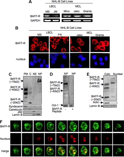
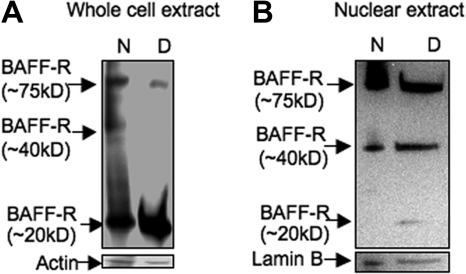
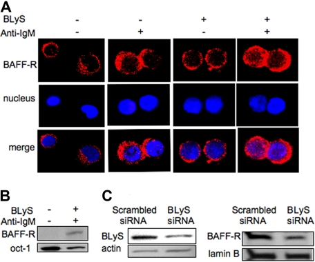
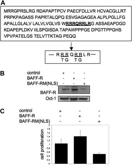
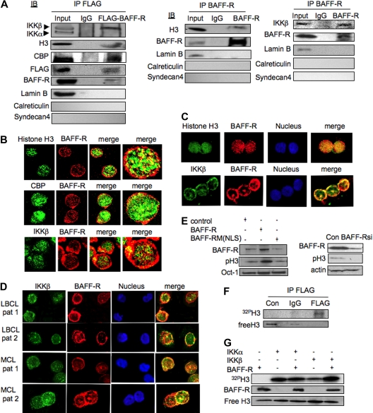
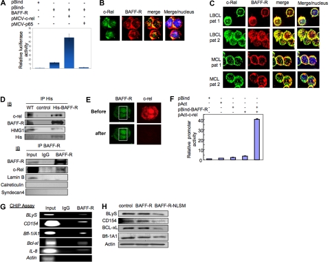
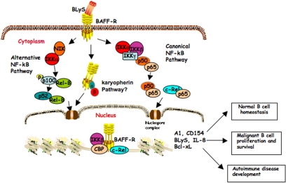
BLyS and its major receptor BAFF-R have been shown to be critical for development and homeostasis of normal B lymphocytes, and for cell growth and survival of neoplastic B lymphocytes, but the biologic mechanisms of this ligand/receptor-derived intracellular signaling pathway(s) have not been completely defined. We have discovered that the BAFF-R protein was present in the cell nucleus, in addition to its integral presence in the plasma membrane and cytoplasm, in both normal and neoplastic B cells. BAFF-R interacted with histone H3 and IKKbeta in the cell nucleus, enhancing histone H3 phosphorylation through IKKbeta. Nuclear BAFF-R was also associated with NF-kappaB/c-Rel and bound to NF-kappaB targeted promoters including BLyS, CD154, Bcl-xL, IL-8, and Bfl-1/A1, promoting the transcription of these genes. These observations suggested that in addition to activating NF-kappaB pathways in the plasma membrane, BAFF-R also promotes normal B-cell and B-cell non-Hodgkin lymphoma (NHL-B) survival and proliferation by functioning as a transcriptional regulator through a chromatin remodeling mechanism(s) and NF-kappaB association. Our studies provide an expanded conceptual view of the BAFF-R signaling, which should contribute a better understanding of the physiologic mechanisms involved in normal B-cell survival and growth, as well as in the pathophysiology of aggressive B-cell malignancies and autoimmune diseases.


Figures

Figure 1
Figure 1
BAFF-R is present in different subcellular compartments in NHL-B cells. (A) RT-PCR analysis of BAFF-R mRNA expression in NHL-B-cell lines. (B) BAFF-R protein was stained for Cy3 (red) fluorescence and a nuclear marker TOPRO-3 (blue) in LBCL and MCL cell lines, and then analyzed by confocal microscopic analysis. (C) Western blot analysis (WB) of protein extract from different subcellular fractions (PM indicates plasma membrane; C, cytoplasm; NE, nuclear envelope; and NP, nucleoplasm) for BAFF-R, syndecan 4 (a plasma membrane marker), calreticulin (an endoplasmic reticulum marker), p62 (a nuclear envelope marker), and lamin B (a nuclear marker). (D) BAFF-R competition experiments were performed by preincubating a BAFF-R peptide with specific BAFF-R antibody before probing nuclear extracts from NHL-B cells. (E) Western blot analysis of 50 μg protein from cytoplasm and nucleoplasm for BAFF-R in normal peripheral blood B cells. Actin and lamin B were used as cytoplasmic and nucleoplasmic markers. (F) Confocal fluorescence microscopic images of GFP-BAFF-R fusion protein localization in a representative live NHL-B cell (MS), cotransfected with BAFF-R expression plasmid pcGFP-BAFF-R (green) and a nuclear localization expression plasmid pDsRed2-Nuc (red).
Figure 2
Figure 2
Nuclear BAFF-R is a high-molecular-weight multimer. (A) Whole-cell lysates (100 μg) from NHL-B cells (MS) transfected with a BAFF-R expression plasmid were treated with (D, denatured) or without (N, nondenatured) β-ME and then probed with anti–BAFF-R antibody by Western blot analysis. (B) Nuclear lysates (50 μg) from NHL-B cells were treated with or without β-ME and then probed with anti–BAFF-R antibody.
Figure 3
Figure 3
BLyS ligand stimulation increases BAFF-R nuclear localization. (A) Normal isolated peripheral blood B cells were stimulated with anti-IgM, BLyS, or both and then probed with BAFF-R (Cy3) and TOPRO-3. Samples were then analyzed by confocal microscopy. (B) WB of nuclear extracts from normal peripheral blood B cells, treated with anti-IgM, human recombinant BLyS, or both, probed with a BAFF-R antibody. Lamin B was used as a nuclear protein loading control. (C) WB of cytoplasm extract from NHL-B cells (MS) transfected with BLyS siRNA (siBLyS) or scrambled siRNA, and probed with BLyS antibody (left panel). Actin was used as loading control. WB of nuclear extracts from NHL-B cells (MS) transfected with BLyS siRNA or scrambled siRNA and probed with BAFF-R antibody. Lamin B was used as a nuclear protein loading control (right panel).
Figure 4
Figure 4
A candidate nuclear localization signal sequence (NLS) detected in BAFF-R is important for NHL-B-cell proliferation and survival. (A) Top is the BAFF-R protein sequence with the putative NLS underlined. Bottom is the wild-type NLS with the mutated amino acids underlined. (B) Nuclear extracts from NHL-B cells transfected with an empty plasmid, a BAFF-R expression plasmid, or a BAFF-R NLS mutant expression plasmid were probed with BAFF-R or Oct-1(nuclear-protein loading control) antibody in WB. (C) A representative 48-hour XTT proliferation assay of NHL-B cells (MS) transfected with a control plasmid, a BAFF-R expression plasmid, or a BAFF-R NLS-mutant expression plasmid was performed. The error bars indicate SD of triplicate samples.
Figure 5
Figure 5
Nuclear BAFF-R interacts with IKKβ and functions in histone H3 phosphorylation. (A) FLAG-BAFF-R fusion protein was purified by FLAG antibody from a nuclear extract of a stably transfected NHL-B-cell line expressing FLAG-BAFF-R. Extract samples were probed with antibodies against CBP, IKKα/β, BAFF-R, or FLAG in WB (left panel). Wild-type BAFF-R was immunoprecipitated (IP) from the nuclear extract using BAFF-R antibody and probed with IKKβ, BAFF-R, or histone H3 antibody on a Western blot (middle and right panels). Lamin B (a nuclear marker), syndecan 4 (a plasma membrane marker), and calreticulin (an endoplasmic reticulum marker) were used as loading controls. (B) Histone H3, CBP, or IKKβ protein in NHL-B cells was identified using Cy2 (green), and BAFF-R protein was identified using Cy3 for confocal microscopic analysis. (C) Histone H3 or IKKβ protein was stained with Cy2, and BAFF-R protein was stained with Cy3 in normal peripheral blood B cells stimulated with anti-IgM and BLyS. Samples were analyzed by confocal microscope. (D) IKKβ protein was stained with Cy2 fluorescence, BAFF-R protein was stained with Cy3, and TOPRO-3 was used as a nuclear marker. (E) Nuclear extracts from NHL-B cells transfected with an empty vector, a BAFF-R expression plasmid, or a BAFF-R NLS-mutant expression plasmid were probed with BAFF-R, phosphorylated histone H3 (Ser10), or Oct-1 (nuclear-protein loading control) antibody in WB (left panel). Whole-cell extracts from NHL-B cells treated with specific BAFF-R siRNA or scrambled control siRNA (control) were probed with BAFF-R, phosphorylated histone H3, or actin antibody in WB (right panel). (F) Recombinant histone H3 was incubated, with or without a FLAG-BAFF-R complex purified by IP with FLAG antibody from NHL-B cells (Jeko) expressing FLAG-BAFF-R protein in an in vitro kinase assay. IgG was used as an IP control. 32P-labeled phosphorylated H3 protein was detected by gel electrophoresis. Unphosphorylated histone H3 (free histone H3) in phosphorylation reaction was detected by WB. (G) Recombinant H3 was incubated with recombinant IKKα or IKKβ, with or without BAFF-R, in an in vitro kinase assay. 32P-labeled phosphorylated H3 was detected by gel electrophoresis. BAFF-R and free histone H3 were detected by WB.
Figure 6
Figure 6
Nuclear BAFF-R interacts with NF-κB/c-Rel and functions as a transcription factor. (A) Comparison of luciferase activity in NHL-B (LBCL-MS) cells transfected with a pBind control vector, a pBindBAFF-R vector, both pBindBAFF-R and c-Rel expression vectors, or both pBindBAFF-R and p65 expression vectors. The error bars indicate SD of triplicate samples. (B) c-Rel protein was stained with Cy2 fluorescence; BAFF-R protein was stained with Cy3 fluorescence in the NHL-B-cell line (MS). Samples were analyzed by confocal microscope. TOPRO-3 was used as a nuclear marker. (C) The above experiment was repeated in representative biopsy-derived LBCL and MCL patient samples, as shown. (D) his-BAFF-R fusion protein was purified by MagneHis purification system from whole-cell lysates of NHL-B cells (MS) transfected with a pcDNA3.1-his-BAFF-R expression plasmid. Samples were probed with BAFF-R antibody, c-Rel antibody, HMG1 antibody, and his antibody (top panel) in WB. Wild-type BAFF-R protein complex was precipitated by BAFF-R antibody from the nuclear extract of NHL-B cells (MS) and probed with BAFF-R or c-Rel antibody in WB (bottom panel). Lamin B (a nuclear marker), syndecan 4 (a plasma membrane marker), and calreticulin (an endoplasmic reticulum marker) were used as loading control. (E) FRET analysis was performed in NHL-B cells (MS) cotransfected with GFP-BAFF-R fusion protein expression plasmid and dsRed–c-Rel fusion protein expression plasmid. Images of a representative transfected cell were taken with a confocal microscope before photobleaching (labeled “pre”) and after photobleaching (labeled “post”). (F) The pACT-c-Rel and pBIND-BAFF-R fusion vectors were cotransfected with pG5luc into NHL-B cells. Firefly and luciferase activity was measured and normalized by β-gal luciferase activity. The error bars indicate SD of triplicate samples. (G) ChIP analysis was performed in NHL-B (LBCL-MS) cells after precipitation of the protein-DNA complex with BAFF-R antibody. PCR analysis to detect the BLyS, CD154, Bfl-1/A1, Bcl-xL, and IL-8 promoters was performed using immunoprecipitated DNA, using proximal primers near the c-Rel binding region in these gene promoters. Actin was used as a control. (H) Whole-cell lysates from NHL-B cells transfected with control (empty) plasmid, a BAFF-R expression plasmid, or a BAFF-R NLS-mutant expression plasmid were blotted with BLyS, CD154, BCL-xL, Bfl-1/A1, or actin (loading control) antibody in WB.
Figure 7
Figure 7
Schematic model of BLyS/BAFF-R signaling in normal and neoplastic B cells. Our current conceptual hypothesis of how the BLyS/BAFF-R signaling contributes to autonomous cell growth in normal and aggressive NHL-B cells. In this model, BAFF-R located in the plasma membrane of lymphoma cells provides signaling for NF-κB pathway activation upon ligand (BLyS) binding. In the nucleus, BAFF-R associates with IKKβ and promotes histone H3 phosphorylation. BAFF-R, IKKβ, and NF-κB/c-rel form a nuclear complex binding to NF-κB target gene promoters and regulating transcription of these genes.

Similar articles

Cited by

References

    1. Yan M, Brady JR, Chan B, et al. Identification of a novel receptor for B lymphocyte stimulator that is mutated in a mouse strain with severe B cell deficiency. Curr Biol. 2001;11:1547–1552. - PubMed
    1. He B, Chadburn A, Jou E, Schattner EJ, Knowles DM, Cerutti A. Lymphoma B cells evade apoptosis through the TNF family members BAFF/BLyS and APRIL. J Immunol. 2004;172:3268–3279. - PubMed
    1. Claudio E, Brown K, Park S, Wang H, Siebenlist U. BAFF-induced NEMO-independent processing of NF-kappa B2 in maturing B cells. Nat Immunol. 2002;3:958–965. - PubMed
    1. Kayagaki N, Yan M, Seshasayee D, et al. BAFF/BLyS receptor 3 binds the B cell survival factor BAFF ligand through a discrete surface loop and promotes processing of NF-kappaB2. Immunity. 2002;17:515–524. - PubMed
    1. Do RK, Hatada E, Lee H, Tourigny MR, Hilbert D, Chen-Kiang S. Attenuation of apoptosis underlies B lymphocyte stimulator enhancement of humoral immune response. J Exp Med. 2000;192:953–964. - PMC - PubMed

Publication types

MeSH terms