. 2009 Mar-Apr;2(3-4):168-77.
doi: 10.1242/dmm.001263. Epub 2009 Feb 2.

Tissue-specific conditional CCM2 knockout mice establish the essential role of endothelial CCM2 in angiogenesis: implications for human cerebral cavernous malformations

Affiliations

Tissue-specific conditional CCM2 knockout mice establish the essential role of endothelial CCM2 in angiogenesis: implications for human cerebral cavernous malformations

Gwénola Boulday et al. Dis Model Mech. 2009 Mar-Apr.

Abstract

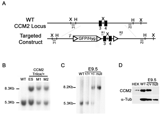
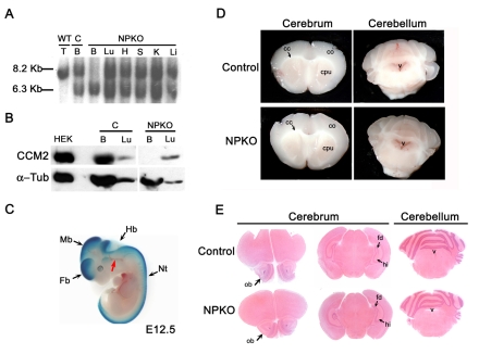
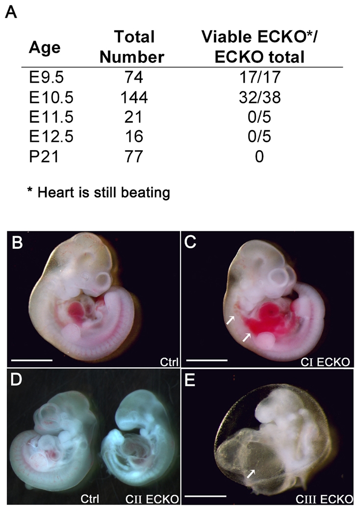
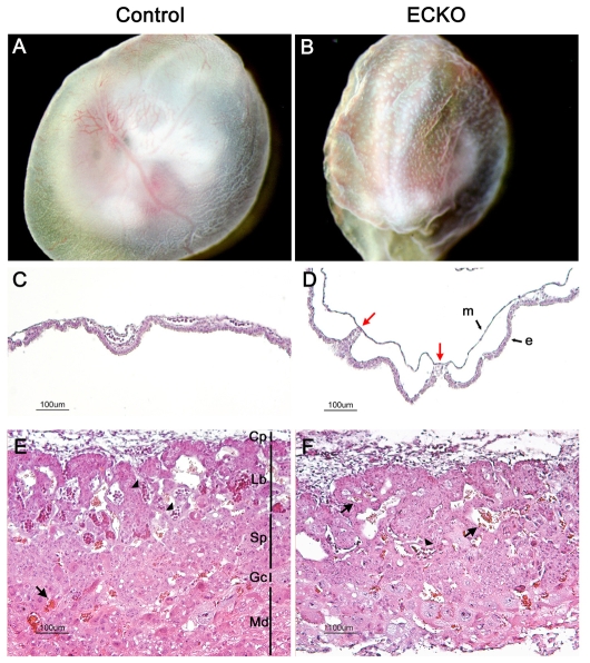
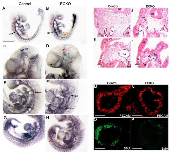
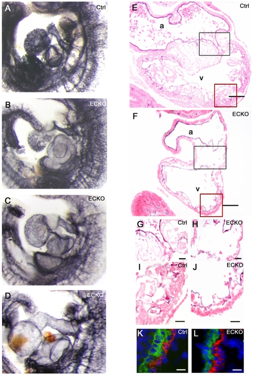
Cerebral cavernous malformations (CCM) are vascular malformations of the brain that lead to cerebral hemorrhages. In 20% of CCM patients, this results from an autosomal dominant condition caused by loss-of-function mutations in one of the three CCM genes. High expression levels of the CCM genes in the neuroepithelium indicate that CCM lesions might be caused by a loss of function of these genes in neural cells rather than in vascular cells. However, their in vivo function, particularly during cerebral angiogenesis, is totally unknown. We developed mice with constitutive and tissue-specific CCM2 deletions to investigate CCM2 function in vivo. Constitutive deletion of CCM2 leads to early embryonic death. Deletion of CCM2 from neuroglial precursor cells does not lead to cerebrovascular defects, whereas CCM2 is required in endothelial cells for proper vascular development. Deletion of CCM2 from endothelial cells severely affects angiogenesis, leading to morphogenic defects in the major arterial and venous blood vessels and in the heart, and results in embryonic lethality at mid-gestation. These findings establish the essential role of endothelial CCM2 for proper vascular development and strongly suggest that the endothelial cell is the primary target in the cascade of events leading from CCM2 mutations to CCM cerebrovascular lesions.


Figures

Fig. 1.
Fig. 1.
Targeted disruption of the mouse Ccm2 gene. (A) Schematic drawing of the Ccm2 gene targeting strategy. The Ccm2 locus was targeted by homologous recombination using a construct containing three loxP sites (open triangles) flanking a hygromycin resistance cassette and Ccm2 exons 3 and 4 (black boxes). The relevant restriction sites (X and H), primers for genotyping (F, forward; R, reverse) and probes for Southern blotting (P1 and P2) are indicated. (B,C) Validation of Ccm2 targeting by Southern blot analysis. 10 μg of genomic DNA from cells, mouse tissues or embryos (as indicated) was digested with XbaI and hybridized with the 5′ external probe, P1. (B) Identification of the ES clone and mice (M1 and M2) containing the targeted Ccm2 allele (indicated as the trilox allele). The 5.3 kb and the 8.3 kb DNA fragments represent the wild-type Ccm2 and targeted alleles, respectively. (C) Southern blot analysis for Ccm2 in either wild-type animals (WT), or wild-type embryos (+/+), heterozygous Ccm2+/– embryos (+/–) or homozygous deleted embryos (null). The 5.3 kb and the 8.2 kb DNA fragments represent the wild-type Ccm2 and the deleted alleles, respectively. (D) Western blot analysis of CCM2 protein expression using 15 μg of total protein lysates from E9.5 embryos (Ccm2+/+ and Ccm2-null embryos). Protein lysate from HEK cells transfected with the Ccm2 cDNA was used as a positive control (200 ng). Immunoblotting for α-tubulin on the same blot was performed as a control for the amount of protein loaded. F, forward primer; H, HindIII; Hyg, hygromycine; P, probe; R, reverse primer; X, XbaI.
Fig. 2.
Fig. 2.
Conditional deletion of CCM2 from neuroglial precursors does not lead to major cerebrovascular defects. (A-C) Analysis of the specific inactivation of CCM2 in the neuroglial compartment. (A) Southern blot analysis using 12 μg of genomic DNA extracted from tissues at P8. DNA was digested by HindIII and hybridized with the external radiolabeled 3′ probe P2. The floxed allele (8.2 kb) in NPKO was recombined specifically within the brain of NPKO mice. A Ccm2–/flox brain from a littermate was used as a control for the absence of recombination (second lane from the left). The 6.3 kb and the 8.2 kb DNA fragments represent the Ccm2-deleted and floxed alleles, respectively. Note that the DNA fragment in the first lane is slightly lower than the floxed fragment and corresponds to the 8.13 kb wild-type allele. (B) Western blot analysis of CCM2 protein expression within the brain (100 μg protein lysates) and the lung (70 μg protein lysates) from NPKO mice and a control littermate at P8. CCM2 protein was not detected in NPKO brain lysates. Protein lysate from Ccm2-transfected HEK cells was used as a positive control (200 ng). Note that the second lane of the blot is free of sample. Immunoblotting for α-tubulin on the same blot was performed as a loading control. (C) β-galactosidase expression analysis on a E12.5 embryo obtained after crossing a nestin-Cre; Ccm2 floxed animal with a Rosa26R reporter line (genotype of the embryo: nestin-Cre; Rosa26R; Ccm2+/flox). Note that the blood vessels (red arrow) are not blue, demonstrating an absence of recombination in the ECs. (D,E) Analysis of the brains from NPKO and control mice. (D) Analysis of 2 mm-thick brain coronal sections from 2-month-old NPKO and control animals under a dissecting microscope, showing slices of the cerebrum (left panels) and the cerebellum (right panels). (E) Hematoxylin and eosin (H&E) staining on 10 μm paraffin-embedded brain sections from NPKO or control mice at P19. B, brain; C, control; cc, corpus callosum; co, cortex; cpu, caudate putamen; Fb, forebrain; fd, fascia dentata; H, heart; Hb, hindbrain; hi, hippocampus; K, kidney; Li, liver; Lu, lung; Mb, midbrain; Nt, neural tube; ob, olfactory bulb; S, spleen; T, toe; v, ventricle.
Fig. 3.
Fig. 3.
Conditional deletion of CCM2 from endothelial cells leads to a variable phenotype at E10.5 and finally to death. (A) ECKO progeny resulting from intercrosses between Tie2-Cre; Ccm2+/– and homozygous Ccm2 floxed mice. Viability was assessed by the presence of heartbeats. At E11.5 and E12.5, no ECKO embryos were found alive (genotyping was performed on embryos under resorption). (B-E) ECKO embryo classes reflecting the severity of the phenotype at E10.5. (B) Control embryo. (C) Class I ECKO embryo (CI) without developmental delay. Note the hemorrhage in the trunk (white arrows). (D) Class II ECKO embryo (CII, right) with a control littermate (left). (E) Class III ECKO embryo (CIII) showing failure to complete turning and an enormous heart with a massively enlarged atrium (white arrow). Note that this embryo shows signs of resorption. Bars, 1 mm (B,C); 500 μm (E).
Fig. 4.
Fig. 4.
Vascular defects in extra-embyonic tissues in ECKO embryos at E10.5. (A,B) Newly dissected E10.5 ECKO YSs are easily distinguishable from control ones owing to their wrinkled surface and their immature vasculature, which remains in a honeycomb pattern. (C,D) H&E staining of E10.5 YS cross sections. Compared with the control YSs (C), the endodermal (e) and mesodermal (m) layers are rarely connected in the ECKO YSs [red arrows in panel (D)]. (E,F) Placenta histology in control (E) or ECKO (F) embryos at E10.5. Note the rare embryonic nucleated red blood cells (arrowheads) in the labyrinthine layer in the ECKO placenta, and the presence of maternal red blood cells (arrows). Cp, chorionic plate; Gc, giant cells; Lb, labyrinthine trophoblaste; Md, maternal decidua; Sp, spongiotrophoblast.
Fig. 5.
Fig. 5.
Conditional deletion of CCM2 from endothelial cells results in major vascular defects in ECKO embryos. (A-H) Whole-mount PECAM staining of control (left panels) and ECKO embryos (right panels) at E9.5 (A,B) and E10.5 (C-H). (I-L) E10.5 PECAM-stained embryo sections after counterstaining with eosin. The presence of PECAM-positive DA, somitic vessels and branchial arch arteries (numbered in red) at E9.5 demonstrates the occurrence of vasculogenesis (A,B). However, at E10.5 (C-H), the arterial and venous blood vessels are abnormal in ECKO embryos and the internal carotid artery is difficult to distinguish [red arrow (D)]. Further, DA are highly irregular in appearance and common cardinal veins are enlarged (F,J). In some animals, the caudal part of the DA fails to fuse (red arrows (H,L)]. (M-P) Double staining for PECAM [red (M,N)] and SMA [green (O,P)] on E10.5 DA sections from the trunk. Note the impaired recruitment of vascular smooth muscle cells/pericytes in the DA of ECKO embryos (P). CCV, common cardinal vein; DA, dorsal aorta; ICA, internal carotid artery; LB, limb bud; NT, neural tube; S, somite; Bars, 500 μm (A,B); 50 μm (M-P).
Fig. 6.
Fig. 6.
Cardiac defects in ECKO embryos. (A-D) Hearts from control (A) and ECKO embryos (B-D) after whole-mount PECAM staining at E10.5. (E-J) Heart sections from PECAM-stained control (E) and ECKO (F) embryos, counterstained with eosin, showing the common atrial chamber (a) and the common ventricular chamber (v). The black and red boxes in (E,F) are shown enlarged in (G,H) and (I,J), respectively. (G,H) A reduction of cells was observed in the atrioventricular canal in the ECKO heart. (I,J) Ventricular trabeculations are strongly reduced in the ECKO heart (J). (K,L) Double staining for PECAM (red) and SMA (green) showing the atrial wall from a control (K) and an ECKO (L) embryo. Sections were counterstained with DAPI. Bars, 100 μm (E,F); 25 μm (G-J); 20 μm (K,L).

References

    1. Akers AL, Johnson E, Steinberg GK, Zabramski JM, Marchuk DA. (2008). Biallelic somatic and germline mutations in cerebral cavernous malformations (CCM): evidence for a two-hit mechanism of CCM pathogenesis. Hum. Mol. Genet. December 16 [Epub ahead of print] [doi:10.1093/hmg/ddn430]. - PMC - PubMed
    1. Bergametti F, Denier C, Labauge P, Arnoult M, Boetto S, Clanet M, Coubes P, Echenne B, Ibrahim R, Irthum B, et al. (2005). Mutations within the programmed cell death 10 gene cause cerebral cavernous malformations. Am. J. Hum. Genet. 76, 42–51 - PMC - PubMed
    1. Brouillard P, Boon LM, Mulliken JB, Enjolras O, Ghassibe M, Warman ML, Tan OT, Olsen BR, Vikkula M. (2002). Mutations in a novel factor, glomulin, are responsible for glomuvenous malformations (“glomangiomas”). Am. J. Hum. Genet. 70, 866–874 - PMC - PubMed
    1. Carmeliet P. (2000). Mechanisms of angiogenesis and arteriogenesis. Nat. Med. 6, 389–395 - PubMed
    1. Clatterbuck RE, Eberhart CG, Crain BJ, Rigamonti D. (2001). Ultrastructural and immunocytochemical evidence that an incompetent blood-brain barrier is related to the pathophysiology of cavernous malformations. J. Neurol. Neurosurg. Psychiatr. 71, 188–192 - PMC - PubMed

Publication types

Substances