. 2009 May 1;107(1):144-54.
doi: 10.1002/jcb.22108.

The osteogenic transcription factor Runx2 regulates components of the fibroblast growth factor/proteoglycan signaling axis in osteoblasts

Affiliations

The osteogenic transcription factor Runx2 regulates components of the fibroblast growth factor/proteoglycan signaling axis in osteoblasts

Nadiya M Teplyuk et al. J Cell Biochem. .

Abstract

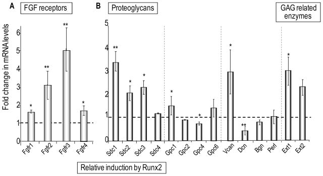
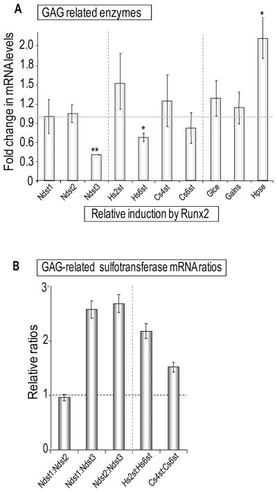
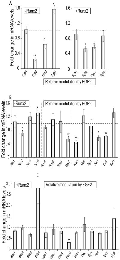
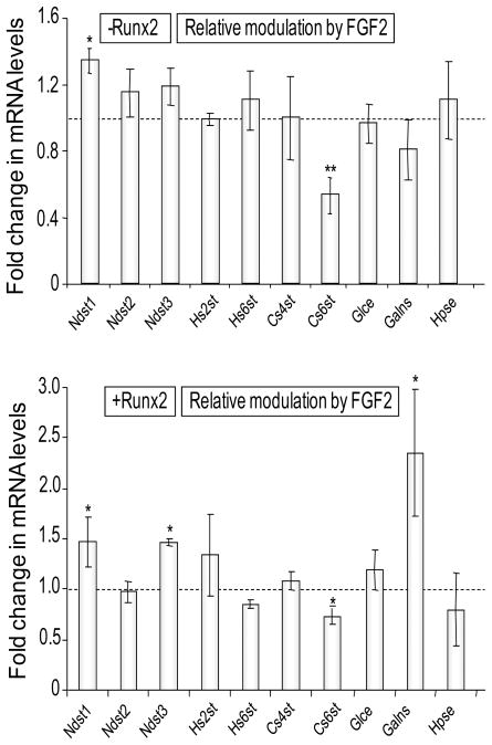
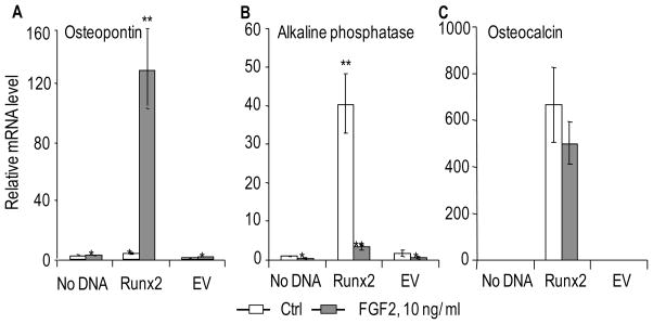
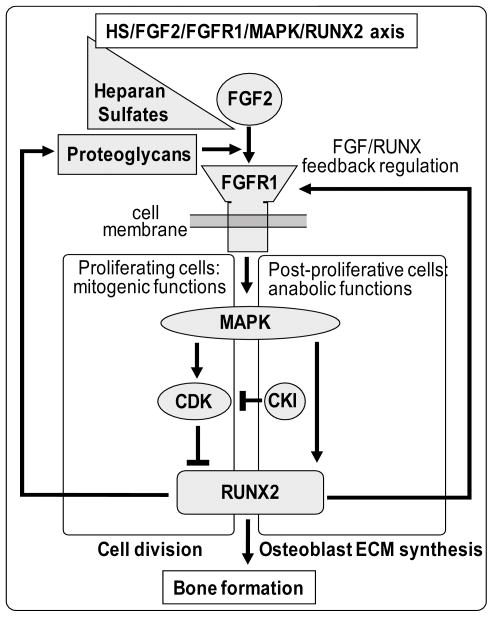
Heparan sulfate proteoglycans cooperate with basic fibroblast growth factor (bFGF/FGF2) signaling to control osteoblast growth and differentiation, as well as metabolic functions of osteoblasts. FGF2 signaling modulates the expression and activity of Runt-related transcription factor 2 (Runx2/Cbfa1), a key regulator of osteoblast proliferation and maturation. Here, we have characterized novel Runx2 target genes in osteoprogenitors under conditions that promote growth arrest while not yet permitting sustained phenotypic maturation. Runx2 enhances expression of genes related to proteoglycan-mediated signaling, including FGF receptors (e.g., FGFR2 and FGFR3) and proteoglycans (e.g., syndecans [Sdc1, Sdc2, Sdc3], glypicans [Gpc1], versican [Vcan]). Runx2 increases expression of the glycosyltransferase Exostosin-1 (Ext1) and heparanase, as well as alters the relative expression of N-linked sulfotransferases (Ndst1 = Ndst2 > Ndst3) and enzymes mediating O-linked sulfation of heparan sulfate (Hs2st > Hs6st) or chondroitin sulfate (Cs4st > Cs6st). Runx2 cooperates with FGF2 to induce expression of Sdc4 and the sulfatase Galns, but Runx2 and FGF2 suppress Gpc6, thus suggesting intricate Runx2 and FGF2 dependent changes in proteoglycan utilization. One functional consequence of Runx2 mediated modulations in proteoglycan-related gene expression is a change in the responsiveness of bone markers to FGF2 stimulation. Runx2 and FGF2 synergistically enhance osteopontin expression (>100 fold), while FGF2 blocks Runx2 induction of alkaline phosphatase. Our data suggest that Runx2 and the FGF/proteoglycan axis may form an extracellular matrix (ECM)-related regulatory feed-back loop that controls osteoblast proliferation and execution of the osteogenic program.


Figures

Figure 1
Figure 1. Runx2 and FGF2 signaling control both proliferation and maturation of osteoblast
A. The schematic depicts the dual role of Runx2 and FGF2 signaling in control of osteoblast proliferation and differentiation. Runx2 may act opposite to FGF2 signaling during the G1 phase of the cell cycle in osteoprogenitors. FGF2 has potent mitogenic effects and stimulates the proliferative expansion of osteoprogenitor cells, while Runx2 suppresses cell cycle progression and may drive cells into quiescence. Runx2 and FGF2 signaling work synergistically on the early steps of osteoblast differentiation, both inducing expression of the early osteoblastic markers (e.g. Osteopontin). B. The experimental system used in this study is a complementation assay in which Runx2 protein is exogenously expressed in osteoprogenitor cells with a homozygous Runx2 null background. Runx2 protein levels were detected by western blot analysis 36 h after infection with recombinant adenovirus expressing Runx2 and GFP (lane ‘Runx2’), after infection with the same vector lacking Runx2 but containing GFP (abbreviated EV; lane ‘Control EV’) or in mock-infected cells (lane ‘No DNA’). CDK2 protein levels were used as internal endogenous control. C. Re-introduction of Runx2 protein expression in Runx2 null cells decreases proliferation as monitored by cell counting (left bar graph) and increased osteogenic differentiation as assessed by Alkaline phosphatase (AP) staining (right bar graph) after cells were grown for 8 days in osteogenic media (50 μg/ml Ascorbic acid, 10 mM β-Glycerophosphate) and treated with 200 ng/ml BMP2 for 24 h. AP staining was quantified by densitometry using ImageJ software. Error bars represent standard error of mean (SE) between three different quantifications. D. Runx2 regulates distinct expression programs. Runx2 regulation of ECM proteins, G protein signaling and sterol metabolism in osteoprogenitor cells were identified previously by Affymetrix gene expression profiling [Teplyuk et al., 2008]. This study focuses on Runx2 regulation of proteoglycan related gene. E. Membrane and extracellular matrix related proteoglycans were identified as a component of Runx2 responsive programs by hierarchical clustering, as well as the DAVID 2.0 database (Database for Annotation, Visualization and Integrated Discovery, http://david.abcc.ncifcrf.gov) [Dennis, Jr. et al., 2003] and information Hyperlinked Over Proteins (iHOP, http://www.ihop-net.org) [Hoffmann and Valencia, 2004].
Figure 2
Figure 2. Relative basal expression levels of genes encoding proteoglycans, FGF receptors and proteoglycan modifying enzymes in osteoprogenitor cells
We determined basal mRNA expression by qPCR analysis in Runx2 null osteoprogenitor cells to assess the relative expression of selected genes in the absence of Runx2 and to determine which genes are characteristic for immature cells within the early osteogenic lineage. The mRNA levels of different genes were plotted as a percentage of GAPDH mRNA levels. Genes were arbitrarily divided based on their expression level into robustly expressed (A) and weakly expressed (B) genes, with the dividing point at 1% of GAPDH expression. Error bars represented Standard error of mean (SE) between three different populations of cells.
Figure 3
Figure 3. Responsiveness of FGF receptors and proteoglycans gene expression by Runx2 in osteoprogenitors
Expression levels of FGF receptors (A) or proteoglycans (B) were determined by qPCR analysis in Runx2 null osteoprogenitors infected with vectors that either do or do not express Runx2 protein. Relative mRNA levels were plotted as fold change of exogenous Runx2 expressing cells over the GFP expressing control and normalized to 18S ribosomal RNA level. Statistical significance of differences was determined by Student’s T-test. Values with P < 0.05 are indicated by asterisks and values with P < 0.01 have two asterisks. Error bars represented Standard error of mean (SE) between three independent experiments. Panel B (right part) also contains data for two glycosaminoglycan (GAG) modifying enzymes that did not fit the scale of the graphs in Figure 4A.
Figure 4
Figure 4. Regulation of glycosaminoglycan modifying enzymes gene expression by Runx2 in osteoprogenitors
Fold changes in mRNA expression of genes encoding glycosaminoglycan (GAG) modifying enzymes including different heparan sulfate and chondroitin sulfate sulfotransferases species upon expression of Runx2 (A), or the same data as in Panel B plotted in a manner that emphasizes relative changes in the expression ratios of enzymes involved in N- versus O-linked modifications of GAGs (B). Similar to Fig. 3, relative mRNA levels were normalized to 18S ribosomal RNA levels and plotted as fold change upon exogenous Runx2 expression in Runx2 null cells. Error bars represent SE between three independent experiments. Statistical significance of differences was determined by Student’s T-test. Values with P < 0.05 are indicated by asterisks and values with P < 0.01 have two asterisks. Error bars represented Standard error of mean (SE) between three independent experiments. Data on expression of two glycosyltransferases (Ext 1 and Ext2) that did not fit the scale of the graph in Fig. 4A were included in Fig. 3B.
Figure 5
Figure 5. FGF2 responsiveness of genes encoding FGF receptors and Proteoglycans in the absence or presence of Runx2
Cells infected with an Adenoviral vector expressing Runx2 or the corresponding empty vector were treated 24 h after infection with 10 ng/ml of bovine FGF2 in DMSO or a corresponding amount of DMSO control for the next 24 h. Relative mRNA expression levels of genes encoding FGF receptors (A) and Proteoglycans (B) were detected by qPCR analysis. The mRNA levels were normalized to 18S ribosomal RNA and plotted as fold change after FGF2 treatment versus DMSO control in the absence (left panel in A, upper panel in B) or in the presence (right panel in A and lower panel in B) of Runx2 expression. Statistical significance of differences was determined by Student’s T-test and values with P < 0.05 are indicated by asterisks, and values with P < 0.01 have two asterisks. Error bars represented Standard error of mean (SE) between three independent experiments.
Figure 6
Figure 6. FGF2 responsiveness of genes encoding glycosaminoglycan modifying enzymes in the absence or presence of Runx2
Similar to Figure 5, cells infected with an Adenoviral vector expressing Runx2 or the corresponding empty vector were treated 24 h after infection with 10 ng/ml of bovine FGF2 in DMSO or a corresponding amount of DMSO control for the next 24 h. Relative mRNA expression levels of genes encoding glycosaminoglycan modifying enzymes were detected by qPCR analysis. The mRNA levels were normalized to 18S ribosomal RNA and plotted as fold change after FGF2 treatment versus DMSO control in the absence (upper panel) or in the presence (lower panel) of Runx2 expression. Statistical significance of differences was determined by Student T-test and values with P < 0.05 are indicated by asterisks, and values with P < 0.01 have two asterisks. Error bars represented Standard error of mean (SE) between three independent experiments.
Figure 7
Figure 7. FGF2 signaling synergizes with Runx2 for the induction of Osteopontin, but antagonizes the induction of Alkaline Phosphatase in osteoprogenitor cells
Similar to Figure 5, cells infected with an Adenoviral vector expressing Runx2 or the corresponding empty vector were treated 24 h after infection with 10 ng/ml of bovine FGF2 in DMSO or a corresponding amount of DMSO control for the next 24 h. Expression of the osteoblastic markers Osteopontin (A), Alkaline phosphatase (B) and Osteocalcin (C) was determined by qPCR analysis. The mRNA levels for the three genes were normalized using 18S ribosomal RNA and plotted relative to the value of Osteocalcin mRNA in the control sample (no infection, DMSO treated) which was arbitrarily set as 1. Statistical significance of differences was determined by Student’s T-test and values with P < 0.05 are indicated by asterisks, and values with P < 0.01 have two asterisks. Error bars represented Standard error of mean (SE) between three independent experiments.
Figure 8
Figure 8. Cross-talk between the Runx2 and FGF2 signaling axes during osteoblastic lineage progression
The model depicts several well-known aspects of the FGF signaling cascade including the synergy between FGF signaling and proteoglycans, as well as downstream effects on FGF signaling on MAPK and CDK related pathways. We propose that Runx2 may participate in two distinct feed-back loops. In actively dividing cells, FGF2 is mitogenic and activates MAPKs and CDKs. This activation may both promote and attenuate Runx2 activity to generate short-term changes in proteoglycan expression that transiently modulate responsiveness to FGF2. In post-proliferative cells, FGF2 functions anabolically and CDK effects on Runx2 are blocked by CDK inhibitors (CKIs). Consequently, the FGF/MAPK/Runx2 pathway may generate a long-term sustained response in which Runx2 modulates a program of proteoglycan expression to promote osteoblast maturation. The ideas presented in the model are consistent with references presented in the main text, but remain to be experimentally tested.

Similar articles

Cited by

References

    1. Bae JS, Gutierrez S, Narla R, Pratap J, Devados R, van Wijnen AJ, Stein JL, Stein GS, Lian JB, Javed A. Reconstitution of Runx2/Cbfa1-null cells identifies a requirement for BMP2 signaling through a Runx2 functional domain during osteoblast differentiation. J Cell Biochem. 2007;100:434–449. - PubMed
    1. Bodo M, Baroni T, Carinci F, Becchetti E, Bellucci C, Conte C, Pezzetti F, Evangelisti R, Tognon M, Carinci P. A regulatory role of fibroblast growth factor in the expression of decorin, biglycan, betaglycan and syndecan in osteoblasts from patients with Crouzon’s syndrome. Eur J Cell Biol. 1999;78:323–330. - PubMed
    1. Bonaventure J, El GV. Molecular and cellular bases of syndromic craniosynostoses. Expert Rev Mol Med. 2003;5:1–17. - PubMed
    1. Boudreaux JM, Towler DA. Synergistic induction of osteocalcin gene expression: identification of a bipartite element conferring fibroblast growth factor 2 and cyclic AMP responsiveness in the rat osteocalcin promoter. J Biol Chem. 1996;271:7508–7515. - PubMed
    1. Coffin JD, Florkiewicz RZ, Neumann J, Mort-Hopkins T, Dorn GW, Lightfoot P, German R, Howles PN, Kier A, O’Toole BA. Abnormal bone growth and selective translational regulation in basic fibroblast growth factor (FGF-2) transgenic mice. Mol Biol Cell. 1995;6:1861–1873. - PMC - PubMed

Publication types

MeSH terms

Substances