

p16(INK4a) immunostaining in cytological and histological specimens from the uterine cervix: a systematic review and meta-analysis
- PMID: 19261387
- PMCID: PMC2784486
- DOI: 10.1016/j.ctrv.2008.10.005
p16(INK4a) immunostaining in cytological and histological specimens from the uterine cervix: a systematic review and meta-analysis
Abstract
Background: P16(INK4a) is a biomarker for transforming HPV infections that could act as an adjunct to current cytological and histological assessment of cervical smears and biopsies, allowing the identification of those women with ambiguous results that require referral to colposcopy and potentially treatment.
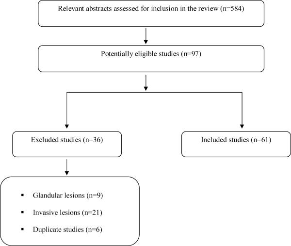
Material and methods: We conducted a systematic review of all studies that evaluated the use of p16(INK4a) in cytological or histological specimens from the uterine cervix. We also estimated the mean proportion of samples that were positive for p16(INK4a) in cytology and histology, stratified by the grade of the lesion.
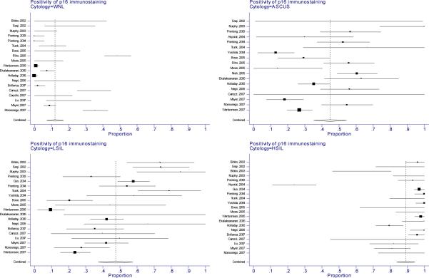
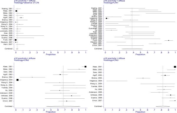
Results: Sixty-one studies were included. The proportion of cervical smears overexpressing p16(INK4a) increased with the severity of cytological abnormality. Among normal smears, only 12% (95% CI: 7-17%) were positive for the biomarker compared to 45% of ASCUS and LSIL (95% CI: 35-54% and 37-57%, respectively) and 89% of HSIL smears (95% CI: 84-95%). Similarly, in histology only 2% of normal biopsies (95% CI: 0.4-30%) and 38% of CIN1 (95% CI: 23-53%) showed diffuse staining for p16(INK4a) compared to 68% of CIN2 (95% CI: 44-92%) and 82% of CIN3 (95% CI: 72-92%).
Conclusion: Although there is good evidence that p16(INK4a) immunostaining correlates with the severity of cytological/histological abnormalities, the reproducibility is limited due to insufficiently standardized interpretation of the immunostaining. Therefore, a consensus needs to be reached regarding the evaluation of p16(INK4a) staining and the biomarker needs to be assessed in various clinical settings addressing specific clinical questions.
Figures
Similar articles
-
Immediate referral to colposcopy versus cytological surveillance for minor cervical cytological abnormalities in the absence of HPV test.Cochrane Database Syst Rev. 2017 Jan 26;1(1):CD009836. doi: 10.1002/14651858.CD009836.pub2. Cochrane Database Syst Rev. 2017. PMID: 28125861 Free PMC article.
-
Role of p16(INK4a) cytology testing as an adjunct to enhance the diagnostic specificity and accuracy in human papillomavirus-positive women within an organized cervical cancer screening program.Acta Cytol. 2012;56(5):506-14. doi: 10.1159/000338979. Epub 2012 Sep 27. Acta Cytol. 2012. PMID: 23075891
-
p16INK4a immunocytochemistry versus human papillomavirus testing for triage of women with minor cytologic abnormalities: a systematic review and meta-analysis.Cancer Cytopathol. 2012 Oct 25;120(5):294-307. doi: 10.1002/cncy.21205. Epub 2012 Jun 14. Cancer Cytopathol. 2012. PMID: 22700382 Free PMC article.
-
Cytology versus HPV testing for cervical cancer screening in the general population.Cochrane Database Syst Rev. 2017 Aug 10;8(8):CD008587. doi: 10.1002/14651858.CD008587.pub2. Cochrane Database Syst Rev. 2017. PMID: 28796882 Free PMC article.
-
[Clinical value of p16INK4a immunocytochemistry in cervical cancer screening].Zhonghua Fu Chan Ke Za Zhi. 2020 Nov 25;55(11):784-790. doi: 10.3760/cma.j.cn112141-20200520-00428. Zhonghua Fu Chan Ke Za Zhi. 2020. PMID: 33228350 Chinese.
Cited by
-
The value of Ki67 for the diagnosis of LSIL and the problems of p16 in the diagnosis of HSIL.Sci Rep. 2022 May 9;12(1):7613. doi: 10.1038/s41598-022-11584-z. Sci Rep. 2022. PMID: 35534530 Free PMC article.
-
The HPV Induced Cancer Resource (THInCR): a Suite of Tools for Investigating HPV-Dependent Human Carcinogenesis.mSphere. 2022 Aug 31;7(4):e0031722. doi: 10.1128/msphere.00317-22. Epub 2022 Aug 11. mSphere. 2022. PMID: 35950764 Free PMC article.
-
High-Risk HPV CISH Detection in Cervical Biopsies with Weak and/or Focal p16 Immunohistochemical Positivity.Int J Mol Sci. 2024 May 14;25(10):5354. doi: 10.3390/ijms25105354. Int J Mol Sci. 2024. PMID: 38791395 Free PMC article.
-
Diagnostic Accuracy of Serum P16ink4A and FOX-P3 Concentrations for Detection of Cervical Lesions Among Women Attending a Cervical Cancer Clinic in Western Uganda: A Case-Control Study.Anal Cell Pathol (Amst). 2025 May 6;2025:1931921. doi: 10.1155/ancp/1931921. eCollection 2025. Anal Cell Pathol (Amst). 2025. PMID: 40365511 Free PMC article.
-
Automated detection of dual p16/Ki67 nuclear immunoreactivity in liquid-based Pap tests for improved cervical cancer risk stratification.Ann Biomed Eng. 2012 May;40(5):1192-204. doi: 10.1007/s10439-011-0498-8. Epub 2012 Jan 4. Ann Biomed Eng. 2012. PMID: 22215277 Free PMC article. Clinical Trial.
References
-
- Ostor AG. Natural history of cervical intraepithelial neoplasia: a critical review. Int J Gynecol Pathol. 1993;12:186–92. - PubMed
-
- Fahey MT, Irwig L, Macaskill P. Meta-analysis of Pap test accuracy. Am J Epidemiol. 1995;141:680–9. - PubMed
-
- Sherman ME, Schiffman MH, Lorinez AT, Manos MM, Scott DR, Kuman RJ, et al. Toward objective quality assurance in cervical cytopathology: Correlation of cytopathologic diagnoses with detection of high-risk human papillomavirus types. Am J Clin Pathol. 1994;102:182–7. - PubMed
-
- Arbyn M, Bergeron C, Klinkhamer P, Martin-Hirsch P, Siebers AG, Bulten J. Liquid compared with conventional cervical cytology. A systematic review and meta-analysis. Obstet Gynecol. 2008;111:167–77. - PubMed
-
- Naucler P, Ryd W, Törnberg S, Strand A, Wadell G, Elfgren K, et al. Human papillomavirus and Papanicolaou tests to screen for cervical cancer. N Engl J Med. 2007;357:1589–97. - PubMed
Publication types
MeSH terms
Substances
Grants and funding
LinkOut - more resources
Full Text Sources
Other Literature Sources
Medical
