

Review
doi: 10.1016/j.ejrad.2009.01.044.
Epub 2009 Mar 3.
Advances in beta-cell imaging
Affiliations
- PMID: 19261414
- PMCID: PMC2680935
- DOI: 10.1016/j.ejrad.2009.01.044
Item in Clipboard
Review
Advances in beta-cell imaging
Eur J Radiol.
2009 May.
Abstract
Diabetes mellitus results in impaired insulin production by pancreatic beta-cells due to their death and/or dysfunction. There is a growing unmet need among diabetes researches and clinicians to assess the level of surviving beta-cells non-invasively. This review will focus on employment of state-of-the-art in vivo imaging methods to estimate and evaluate beta-cell mass in animal models of diabetes.
Figures
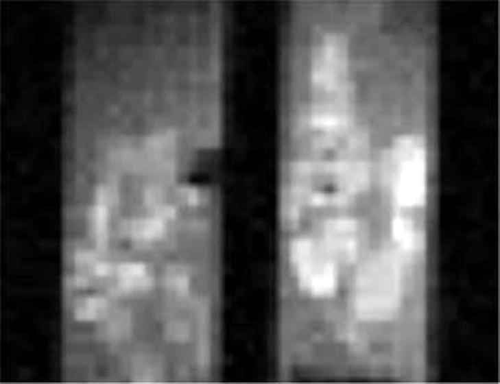
Mn2+-enhanced T1-weighted contrast of rat pancreatic islets. Two capillary tubes containing pancreatic-islets were incubated for 30 minutes in the presence of 25 mM MnCl2. The tubes were incubated in 5 mM glucose (left) and 16.7 mM glucose (right). The image was acquired at 500 MHz with TR = 400 ms, TE = 7.2 ms, slice thickness = 100 mm, field-of-view = 4.8 mm × 2.4mm, acquisition matrix = 128 × 64, and number of averages = 64. Reprinted from Gimi et al. (2006) with kind permission from Cognizant Communication Corp.
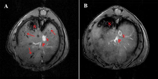
In vivo imaging of intrahepatically transplanted human islets. A: Representative images of NOD.scid mice with transplanted islets. On in vivo images, Feridex-labeled islets appeared as signal voids scattered throughout the liver. B: Non-labeled islets were not detectable using the same imaging parameters. S – stomach, P – portal vein, V – superior vena cava. Reprinted from Evgenov et al. (2006) with kind permission from the American Diabetes Association.
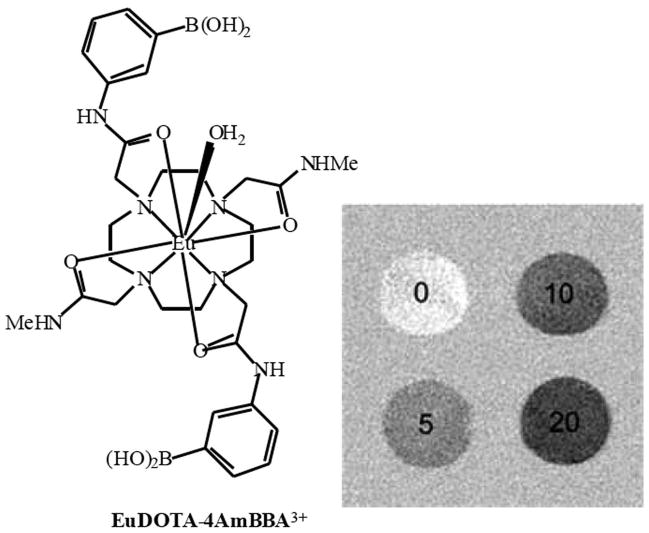
CEST images of phantoms containing 10 mM EuDOTA-4AmBBA plus either 0, 5, 10 or 20 mM glucose, which was obtained by subtracting the image obtained by saturating at 50 ppm from that at 30 ppm. Reprinted from Zhang et al. (2003) with kind permission from the American Chemical Society.
References
-
- Samli K, McGuire M, Newgard C, Johnston S, Brown K. Peptide-mediated targeting of the islets of Langerhans. Diabetes. 2005;54:2103–2108. - PubMed
-
- Hadjivassiliou V, Green M, Green I. Immunomagnetic purification of beta-cells from rat islets of Langerhans. Diabetologia. 2000;43:1170–1177. - PubMed
-
- Hampe C, Wallen A, Schlosser M, Ziegler M, Sweet I. Quantitative evaluation of a monoclonal antibody and its fragment as potential markers for pancreatic beta cell mass. Exp Clin Endocrinol Diabetes. 2005;113:381–387. - PubMed
-
- Brogren C, Hirsch F, Wood P, Poussier P. Production and characterization of a monoclonal islet cell surface autoantibody from the BB rat. Diabetologia. 1986;29:330–333. - PubMed
Publication types
MeSH terms
Grants and funding
LinkOut - more resources
Full Text Sources
Other Literature Sources
Medical
