. 2009 May 1;284(18):11781-91.
doi: 10.1074/jbc.M808703200. Epub 2009 Mar 4.

Ligand-independent thrombopoietin mutant receptor requires cell surface localization for endogenous activity

Affiliations

Ligand-independent thrombopoietin mutant receptor requires cell surface localization for endogenous activity

Caroline Marty et al. J Biol Chem. .

Abstract

The activating W515L mutation in the thrombopoietin receptor (MPL) has been identified in primary myelofibrosis and essential thrombocythemia. MPL belongs to a subset of the cytokine receptor superfamily that requires the JAK2 kinase for signaling. We examined whether the ligand-independent MPL(W515L) mutant could signal intracellularly. Addition of the endoplasmic reticulum (ER) retention KDEL sequence to the receptor C terminus efficiently locked MPL(W515L) within its natural ER/Golgi maturation pathway. In contrast to cells expressing the parental MPL(W515L), MPL(W515L)-KDEL-expressing FDC-P1 cells were unable to grow autonomously and to produce tumors in nude mice. When observed, tumor nodules resulted from in vivo selection of cells leaking the receptor at their surface. JAK2 co-immunoprecipitated with MPL(W515L)-KDEL but was not phosphorylated. We generated disulfide-bonded MPL(W515L) homodimers by the S402C substitution, both in the normal and KDEL context. Unlike MPL(W515L)-KDEL, MPL(W515L-S402C)-KDEL signaled constitutively and exhibited cell surface localization. These data establish that MPL(W515L) with appended JAK2 matures through the ER/Golgi system in an inactive conformation and suggest that the MPL(W515L)/JAK2 complex requires membrane localization for JAK2 phosphorylation, resulting in autonomous receptor signaling.


Figures

FIGURE 1.
FIGURE 1.
Cell surface and total expression of the different MPL constructs in FDC-P1 cells. A, FDC-P1 cells were transduced with the bicistronic retroviral pMX-IRES-CD4 vector encoding either the HA epitope-tagged MPLWT, MPLW515L, or MPLW515L-KDEL. Cells were sorted for equal CD4 levels and either HA-positive or -negative cell surface expression, accordingly, with anti-CD4-APC and anti-HA-PE antibodies. A cell population with MPLW515L-KDEL leaking at the membrane (MPLW515L-KDELleak) due to the saturation of the KDEL retrieval system was selected. Sorted FDC-P1 cell populations were assessed by FACS analysis. B, median PE fluorescence intensities represent the quantities of HA-MPL localized at the cell surface, and AU represents arbitrary units. C, FDC-P1 cell lines expressing each of the MPL constructs were lysed in Laemmli buffer, and cell homogenates were resolved by SDS-PAGE. Expression of the HA-MPL constructs was examined by Western blotting with anti-HA.11 mAb (top panel). Black arrows point to three differentially glycosylated forms of MPL: 95, 80, and 65 kDa for 1, 2 and 3, respectively. The expression of β-actin serves as loading control (bottom panel). Data are representative of similar results obtained in at least three independent experiments.
FIGURE 2.
FIGURE 2.
Cellular localization of the MPLW515L and MPLW515L-KDEL in FDC-P1 cells. Lysates of MPLW515L and MPLW515L-KDEL-expressing FDC-P1 cells were treated with Endo H. The digested (+) and non-digested (–) products corresponding to the differentially glycosylated forms of MPL were detected by Western blotting with anti-HA.11 mAb, as in Fig. 1C. Only the upper band 1 (95kDa) was Endo H-resistant. β-Actin serves as loading control. Blots shown were repeated in three independent experiments.
FIGURE 3.
FIGURE 3.
Proliferation assays of FDC-P1 cells expressing the different MPL constructs. Sorted transduced cell populations expressing each of the MPL constructs were cultured either in absence of cytokine (•), presence of 10 ng/ml hTPO (▵) or 20 ng/ml mIL-3 (□). Viable cells were counted after 2, 3, 4, 7, and 10 days. Results shown reflect averages of triplicate values ± S.D. Results are representative of a typical experiment.
FIGURE 4.
FIGURE 4.
Signaling studies in MPLWT-, MPLW515L-, and MPLW515L-KDEL-expressing FDC-P1 cells. A, cells expressing either MPLWT or MPLW515L were serum- and cytokine-starved for 4 h prior to a 10-min stimulation with 10 ng/ml hTPO at 37 °C, as indicated. Cells were lysed in Laemmli buffer, and the phosphorylation status of JAK2, STAT5, AKT, and ERK1/2 was examined by Western blotting with the respective anti-phospho-specific antibodies, as indicated. Expression of β-actin in the samples was used as loading control. B, similar studies were conducted with parental FDC-P1 cells and cells expressing either MPLW515L or MPLW515L-KDEL stimulated with 10 ng/ml hTPO or 20 ng/ml mIL-3 for 10 min at 37 °C. NS, non-stimulated. Blots shown were reproduced in two independent experiments.
FIGURE 5.
FIGURE 5.
Tumorigenic effect of MPLW515L- and MPLW515L-KDEL-expressing FDC-P1 cells in nude mice. Mice were injected subcutaneously with 4 × 10⁶ of either parental FDC-P1 cells as a negative control (5 mice) or MPLW515L-or MPLW515L-KDEL-expressing cells (10 mice per assay). A, number of animals developing a tumor and surface area of subcutaneous tumors measured in the exponential growth phase (day 12) expressed as mean ± S.D. B, injected cells (day 0) and cells dissociated from two excised tumors at days 18 and 56 were analyzed by FACS with anti-CD4-APC and anti-HA-PE antibodies. Cells were gated on CD4 expression, and cell surface localization of MPL was assessed by using HA.11 mAb labeling.
FIGURE 6.
FIGURE 6.
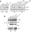
Interaction of JAK2 with MPLWT, MPLW515L, and MPLW515L-KDEL. A, cells expressing either MPLWT or MPLW515L were serum- and cytokine-starved for 4 h and stimulated with 10 ng/ml hTPO for 10 min at 37 °C. Cells were lysed, and HA-tagged MPL constructs were selectively immunoprecipitated using an anti-HA.11 mAb. The immunoprecipitates and cell homogenates (H) were resolved by SDS-PAGE. The ability of JAK2 to associate with the two MPL constructs was examined by Western blotting analysis of the IP with an anti-total JAK2 antibody (IP, middle panel). The phosphorylation status of the bound JAK2 was assessed using an anti-phospho-JAK2 (Tyr-1007/1008) antibody (IP, upper panel). MPL protein in the IP was detected with an anti-MPL antibody and serves as loading control (IP, lower panel). The blot was probed for the expression of JAK2 in H. B, similar experiments were performed with sorted FDC-P1 expressing MPLW515L or MPLW515L-KDEL stimulated with 10 ng/ml hTPO or 20 ng/ml mIL-3 for 10 min at 37 °C. NS, non-stimulated. Results shown are representative of a typical experiment.
FIGURE 7.
FIGURE 7.
Forced dimerization of MPLWT supports cytokine-independent cell growth. A, parental FDC-P1 and MPLWT- and MPLWT-S402C-expressing cells were collected in Laemmli buffer containing or not 2% β-mercaptoethanol (β-Meth). Protein samples were resolved by Western blotting on an acrylamide 4–12% gradient gel (Invitrogen) with anti-HA.11 mAb. The arrow indicates the predicted size of dimeric MPL. B, surface expression of MPLWT and MPLWT-S402C was measured in transduced CD4- and HA-positive sorted FDC-P1 cells with anti-HA-PE antibody by FACS analysis. C, FDC-P1 and cells expressing either MPLWT or MPLWT-S402C were cultured in absence of cytokine (•) or presence of 10 ng/ml hTPO (▵). As a control, FDC-P1 cells were also grown with 20 ng/ml mIL-3 (□). Viable cells were counted after 2, 3, 4, and 7 days. Data are means ± S.D. of triplicate samples and are representative of a typical experiment.
FIGURE 8.
FIGURE 8.
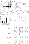
Forced dimerization of MPLW515L-KDEL restores autonomous cell growth. A, cells were lysed in Laemmli buffer containing or not 2% β-mercaptoethanol (β-Meth), and the presence of dimeric (arrow) and monomeric forms of MPL was examined by Western blotting with anti-HA.11 mAb. B, surface expression of MPLW515L, MPLW515L-S402C, MPLW515L-KDEL, and MPLW515L-S402C-KDEL was verified in transduced and sorted FDC-P1 cells with anti-HA-PE antibody and FACS analysis. C, the various cell populations expressing the MPL constructs, as indicated, were cultured either in absence of cytokine (•) or presence of 10 ng/ml hTPO (▵). FDC-P1 cells were grown with 20 ng/ml mIL-3 (□), to serve as control. Viable cells were counted at day 2, 3, 4, and 7. Results presented are means ± S.D. and are representative of a typical experiment. D, median fluorescence intensity of the anti-HA-PE antibody labeling was used for quantification of cell surface expression of MPLW515L-S402S-KDEL at days 0, 3, and 7 of culture in the absence of cytokine (NS). MPLW515L and MPLW515L-KDEL cell surface expressions were also examined. At day 3 MPLW515L-KDEL-expressing cells were dead. As a reference, cells maintained 7 days in the presence of IL-3, thus without selection pressure, were analyzed (IL-3). AU, arbitrary units. E, the Y Log scale of the factor-independent proliferation curve of MPLW515L-S402C-KDEL-expressing cells was converted to linear for the first 3 days. The error bars represent ±S.D. from triplicate samples. F, after cytokine withdrawal (NS), MPLW515L-, MPLW515L-KDEL-, and MPLW515L-S402C-expressing cells were collected at 7, 24, and 48 h, and the nuclei were stained with propidium iodide and analyzed by FACS. FDC-P1 cells grown with IL-3 served as proliferation control (IL-3). Results shown are representative of two independent experiments.
FIGURE 9.
FIGURE 9.
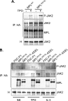
No further TPO-induced JAK2 constitutive activation by MPLW515L carrying the S402C dimerizing mutation. A, FDC-P1 cells expressing the indicated MPL forms were serum- and cytokine-starved for 4 h prior or not (NS) to a 10-min stimulation with 10 ng/ml hTPO at 37 °C (TPO). Cells were lysed in Laemmli buffer, and the phosphorylation status of JAK2 was examined by Western blotting with its anti-phospho specific antibody. Expression of β-actin serves as the loading control. B, MPLW515L- and MPLW515L-S402C-expressing cells were starved and stimulated with TPO as mentioned in A. Cells, including parental FDC-P1 as control, were lysed, and the two HA-tagged receptors were selectively immunoprecipitated with anti-HA.11 mAb. The ability of JAK2 to associate with the receptors was examined by blotting the immunoprecipitates with the anti-JAK2 antibody. The phosphorylation status of the bound JAK2 was assessed by blotting the IP with an anti-phospho-JAK2 (Tyr-1007/1008) antibody. Levels of JAK2 expression were detected in the homogenates (H). MPL in the IP serves as loading control. Blots are representative of similar results obtained in two independent experiments.

Similar articles

Cited by

References

    1. Broudy, V. C., and Kaushansky, K. (1995) J. Leukocyte Biol. 57 719–725 - PubMed
    1. Gurney, A. L., Carver-Moore, K., de Sauvage, F. J., and Moore, M. W. (1994) Science 265 1445–1447 - PubMed
    1. Kaushansky, K., Lok, S., Holly, R. D., Broudy, V. C., Lin, N., Bailey, M. C., Forstrom, J. W., Buddle, M. M., Oort, P. J., Hagen, F. S., Roth, G. J., Papayannopoulou, T., and Foster, D. C. (1994) Nature 369 568–571 - PubMed
    1. Vainchenker, W., Methia, N., Debili, N., Titeux, M., and Wendling, F. (1995) Thromb. Haemostasis 74 526–528 - PubMed
    1. Wendling, F. (1999) Haematologica 84 158–166 - PubMed

Publication types

MeSH terms

LinkOut - more resources