. 2009 Apr 24;284(17):11059-69.
doi: 10.1074/jbc.M806561200. Epub 2009 Mar 4.

Calcitonin, a regulator of the 25-hydroxyvitamin D3 1alpha-hydroxylase gene

Affiliations

Calcitonin, a regulator of the 25-hydroxyvitamin D3 1alpha-hydroxylase gene

Yan Zhong et al. J Biol Chem. .

Abstract

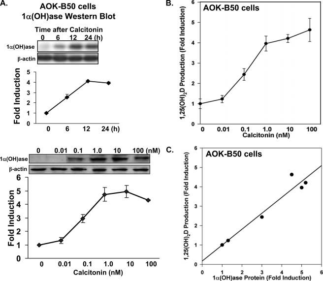
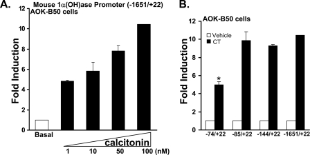
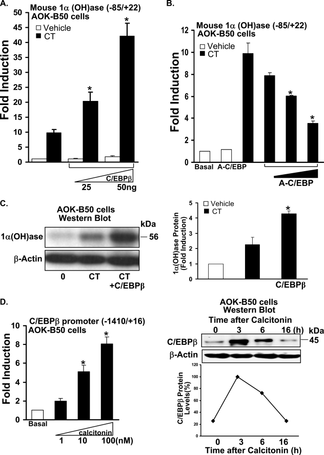
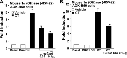
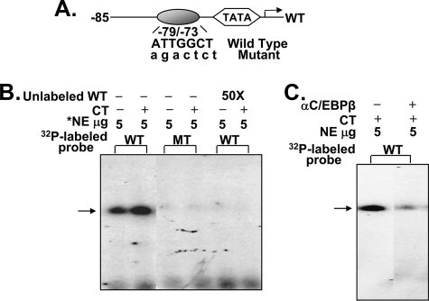
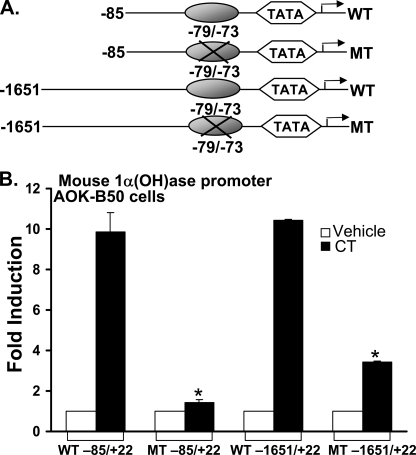
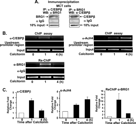
Although parathyroid hormone (PTH) induces 25-hydroxyvitamin D(3) (25(OH)D(3)) 1alpha-hydroxylase (1alpha(OH)ase) under hypocalcemic conditions, previous studies showed that calcitonin, not PTH, has an important role in the maintenance of serum 1,25-dihydroxyvitamin D(3) (1,25(OH)(2)D(3)) under normocalcemic conditions. In this study we report that 1alpha(OH)ase transcription is strongly induced by calcitonin in kidney cells and indicate mechanisms that underlie this regulation. The transcription factor C/EBPbeta is up-regulated by calcitonin in kidney cells and results in a significant enhancement of calcitonin induction of 1alpha(OH)ase transcription and protein expression. Mutation constructs of the 1alpha(OH)ase promoter demonstrate the importance of the C/EBPbeta binding site at -79/-73 for activation of the 1alpha(OH)ase promoter by calcitonin. The SWI/SNF chromatin remodeling complex was found to cooperate with calcitonin in the regulation of 1alpha(OH)ase. Chromatin immunoprecipitation analysis showed that calcitonin recruits C/EBPbeta to the 1alpha(OH)ase promoter, and Re-chromatin immunoprecipitation analysis (sequential chromatin immunoprecipitations using different antibodies) showed that C/EBPbeta and BRG1, an ATPase that is a component of the SWI/SNF complex, bind simultaneously to the 1alpha(OH)ase promoter. These findings are the first to address the dynamics between calcitonin, C/EBPbeta, and SWI/SNF in the regulation of 1alpha(OH)ase and provide a mechanism, for the first time, for calcitonin induction of 1alpha(OH)ase. Because plasma calcitonin as well as 1,25(OH)(2)D(3) have been reported to be increased during pregnancy and lactation and in early development, these findings suggest a mechanism that may account, at least in part, for the increase in plasma 1,25(OH)(2)D(3) during these times of increased calcium requirement.


Figures

FIGURE 1.
FIGURE 1.
Regulation of 1α(OH)ase protein and catalytic activity by calcitonin in AOK-B50 cells. A, Western blot was performed using total extracts from AOK-B50 porcine kidney proximal tubule cells (LLCPK1 cells that express PTH/PTHrP Type 1 receptors as well as calcitonin receptors (31, 32)). Upper panel, cells were treated with vehicle (0) or with calcitonin (100 nm) for 6, 12, or 24 h. Lower panel, AOK-B50 cells were treated with vehicle (0) or increasing concentrations of calcitonin (0.01–100 nm). Results represent the mean ± S.E. of three separate experiments. For all times of calcitonin treatment (A) and for all concentrations of calcitonin (lower panel), 1α(OH)ase levels were significantly induced compared with vehicle (p < 0. 05). B, 1,25(OH)2D3 production in AOK-B50 cells in response to increasing concentrations of calcitonin. Cells were treated with vehicle (0) or with calcitonin (0.01, 0.1, 1, 10, 100 nm) for 12 h and then incubated with 100 nm 25(OH)D3 for 4 h. Cellular 1,25(OH)2D3 production was determined by radioimmunoassay as described under “Experimental Procedures.” Results are reported as the mean ± S.E. (n = 4). Calcitonin treatment (0.1–100 nm) resulted in a significant increase in 1,25(OH)2D3 production (p < 0.05). C, correlation between 1α(OH)ase protein (A, lower panel) and 1,25(OH)2D3 production (B) in AOK-B50 cells in response to calcitonin (r = 0.97, p < 0.01).
FIGURE 2.
FIGURE 2.
A regulatory region for calcitonin stimulation of 1α(OH)ase transcription is localized within –85/+22. A, AOK-B50 cells were plated in a 24-well culture dish, and cells in each well were transfected with 0.3 μg of the mouse 1α(OH)ase promoter construct (–1651/+22). After 24 h, cells were treated with vehicle (Basal) or 1–100 nm calcitonin for another 24 h and harvested, and luciferase activity was determined and normalized based on protein contents of cell lysates. 1α(OH)ase promoter activity is represented as -fold induction (mean ± S.E.; n = 3–6 observations per group) by comparison to basal levels. Calcitonin treatment (1–100 nm) resulted in a significant increase in 1α(OH)ase promoter activity compared with basal levels (p < 0.05). For all transcription experiments, empty vectors were used to keep the total DNA concentration the same. B, AOK-B50 cells were plated in a 24-well culture dish, and cells in each well were transfected with 0.3 μg of mouse 1α(OH)ase promoter –1651/+22 and deletion constructs (–144/+22, –85/+22, –74/+22). After 24 h, cells were treated with vehicle or 100 nm calcitonin (CT) for another 24 h. Results represent the mean ± S.E. of 4–8 observations/group. *, p < 0.05 compared with the activity of the –1651/+22 promoter and the two deletion constructs in response to calcitonin.
FIGURE 3.
FIGURE 3.
Cooperative role of C/EBPβ in the regulation of 1α(OH)ase by calcitonin and calcitonin induction of C/EBPβ protein and transcription in kidney cells. A and B, AOK-B50 cells were plated in a 24-well culture dish, and cells in each well were co-transfected with 0.3 μg of mouse 1α(OH)ase promoter construct (–85/+22) and C/EBPβ expression vector (0.025 and 0.050 μg) (A) or A-C/EBP (0.25, 0.5, 1.0 μg) (B). After 24 h, cells were treated with vehicle or 100 nm calcitonin (CT) for another 24 h. C, C/EBPβ enhances calcitonin induced 1α(OH)ase protein levels. AOK-B50 cells in 100-mm tissue culture dishes were transfected with pMEX-C/EBPβ for 24 h and treated with vehicle (0) or with calcitonin (100 nm) for 6 h (a time when the level of 1α(OH)ase protein induced by calcitonin is suboptimal (see Fig. 1A)). Left panel, representative Western blot. Right panel, graphic representation of densitometric scans of Western blots from three separate experiments (mean ± S.E.). D, calcitonin induces C/EBPβ transcription and protein in AOK B-50 cells. Left panel, AOK-B50 cells were transfected with 0.3 μg of C/EBPβ promoter construct (–1400/+16). After 24 h, cells were treated with vehicle (Basal) or 1–100 nm calcitonin for another 24 h. Calcitonin (10 and 100 nm) significantly induced C/EBPβ promoter activity (p < 0.05). Right panel, top, representative Western blot of C/EBPβ expression in nuclear extracts from AOK-B50 cells treated with vehicle or with calcitonin (100 nm) for 3–16 h. Bottom, graphic representation of densitometric scans of Western blots from three separate experiments (mean ± S.E.). Western blot analysis of nuclear extracts from MCT cells also showed low levels of C/EBPβ at 0 time and induction of C/EBPβ at 3 and 6 h (not shown). 1α(OH)ase (A and B) or C/EBPβ (D, left panel) promoter activity was measured by firefly luciferase activity/protein concentration and represented as -fold induction (mean ± S.E.; n = 3 or more experiments) by comparison to basal levels. *, p < 0.05 compared with calcitonin alone (A, B, and C). *, p < 0.05 compared with basal (D, left panel).
FIGURE 4.
FIGURE 4.
Mutant Brm or BRG1, which act as dominant negative inhibitors, inhibit calcitonin induction of 1α(OH)ase transcription. AOK-B50 cells were co-transfected with 0.3 μg mouse 1α(OH)ase promoter (–85/+22) and Brm-DN expression vectors (0.05, 0.1 μg) (A) or BRG1-DN (0.1 μg) (B). After 24 h, cells were treated with vehicle or 100 nm calcitonin (CT) for another 24 h. 1α(OH)ase promoter activity was measured by firefly luciferase activity/protein concentration and represented as -fold induction (mean ± S.E.; n = at least three observations per group) by comparison to basal levels. Similar results were obtained using the –1651/+22 promoter construct (not shown). Note that there was no effect of 0.1 μg of Brm-DN or 0.1 μg of BRG1-DN on basal levels of 1α(OH)ase transcription (open bar, Brm-DN (A); open bar, BRG1-DN (B)).
FIGURE 5.
FIGURE 5.
C/EBPβ binding site in 1α(OH)ase promoter detected by electrophoretic mobility shift assay. A, schematic of the C/EBPβ binding site at –79/–73 in the mouse 1α(OH)ase promoter. Oligonucleotides corresponding to wild type or mutated C/EBPβ binding site were used. B, calcitonin (CT) treatment (100 nm, 6 h) resulted in increased binding to the C/EBPβ binding site. No binding was observed using the mutated sequence (MT) or preincubation with the unlabeled wild type oligonucleotide (WT). C, preincubation with C/EBPβ antibody depleted the binding. Results are representative of four separate experiments. *, NE, AOK-B50 cell nuclear extract.
FIGURE 6.
FIGURE 6.
Mutation of the C/EBPβ binding site (–79/–73) inhibits the activation of 1α(OH)ase transcription mediated by CT. AOK-B50 cells were transfected with 0.3 μg of mouse 1α(OH)ase (–85/+22) promoter (wild type (WT) or mutant (MT)) or 1α(OH)ase (–1651/+22) promoter (wild type or mutant). After 24 h, cells were treated with vehicle or 100 nm calcitonin (CT) for another 24 h. 1α(OH)ase promoter activity was measured by firefly luciferase activity/protein concentration and represented as -fold induction (mean ± S.E.) by comparison to basal levels (3–5 observations/group). *, p < 0.05 compared with wild type, calcitonin-treated.
FIGURE 7.
FIGURE 7.
C/EBPβ and BRG1 are components of the same nuclear complex and calcitonin modulates C/EBPβ and BRG1 recruitment to the 1α(OH)ase promoter. A, nuclear extracts were prepared from MCT cells and used for immunoprecipitation (IP) with C/EBPβ antibody, BRG1 antibody, or control rabbit IgG. WB, Western blot. B, ChIP analysis of CEBPβ, acetylated histone H4 (AcH4), and Re-ChIP analysis of BRG1 binding to the 1α(OH)ase promoter. MCT cells were treated with vehicle or calcitonin for 1 and 4 h and cross-linked by 1% formaldehyde for 15 min. Cross-linked cell lysates were subjected to immunoprecipitation first with C/EBPβ antibody (α-C/EBPβ) and then with BRG1 antibody (α-BRG1). DNA precipitates were isolated and then subjected to PCR using specific primers designed according to the C/EBPβ site on the mouse 1α(OH)ase promoter (see “Experimental Procedures”). Analysis of input DNA (0.2%) was taken before precipitation (Input). Recruitment of Brm to the 1α(OH)ase promoter was not observed (it should be noted that, although Brm and BRG1 were detected in AOK-B50 cell nuclear extracts, BRG1 but not Brm was detected by Western blot analysis of nuclear extracts of MCT cells). Using a distal 1α(OH)ase promoter region (–1300/–980) binding of C/EBPβ and BRG1 was not observed. C, quantitation of ChIP analyses (±S.E.).

Similar articles

Cited by

References

    1. Bikle, D., Adams, J., and Christakos, S. (2008) in Primer on Metabolic Bone Diseases and Disorder of Mineral Metabolism (Rosen, C., ed) pp. 141–149, American Society for Bone and Mineral Research, Washington DC
    1. Omdahl, J. L., Bobrovnikova, E. V., Annalora, A., Chen, P., and Serda, R. (2003) J. Cell. Biochem. 88, 356–362 - PubMed
    1. Henry, H. (2005) in Vitamin D (Feldman, D., Glorieux, F. H., and Pike, J. W., ed) pp. 69–83, Academic Press, Inc., San Diego, CA
    1. St-Arnaud, R., Messerlian, S., Moir, J. M., Omdahl, J. L., and Glorieux, F. H. (1997) J. Bone Miner. Res. 12, 1552–1559 - PubMed
    1. Kitanaka, S., Takeyama, K., Murayama, A., Sato, T., Okumura, K., Nogami, M., Hasegawa, Y., Niimi, H., Yanagisawa, J., Tanaka, T., and Kato, S. (1998) N. Engl. J. Med. 338, 653–661 - PubMed

Publication types

LinkOut - more resources