. 2009 Apr 24;284(17):11121-33.
doi: 10.1074/jbc.M806268200. Epub 2009 Mar 4.

Bortezomib overcomes tumor necrosis factor-related apoptosis-inducing ligand resistance in hepatocellular carcinoma cells in part through the inhibition of the phosphatidylinositol 3-kinase/Akt pathway

Affiliations

Bortezomib overcomes tumor necrosis factor-related apoptosis-inducing ligand resistance in hepatocellular carcinoma cells in part through the inhibition of the phosphatidylinositol 3-kinase/Akt pathway

Kuen-Feng Chen et al. J Biol Chem. .

Abstract

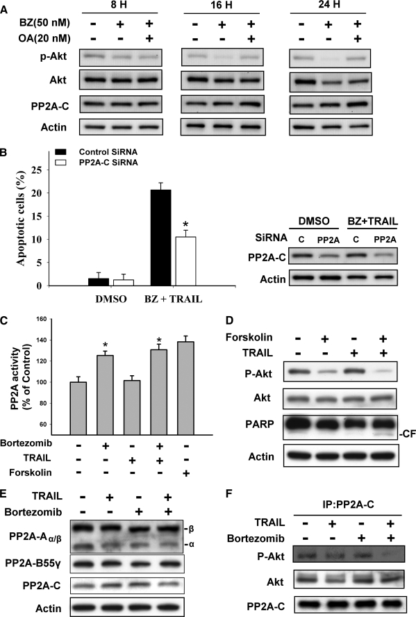
Hepatocellular carcinoma (HCC) is one of the most common and aggressive human malignancies. Recombinant tumor necrosis factor-related apoptosis-inducing ligand (TRAIL) is a promising anti-tumor agent. However, many HCC cells show resistance to TRAIL-induced apoptosis. In this study, we showed that bortezomib, a proteasome inhibitor, overcame TRAIL resistance in HCC cells, including Huh-7, Hep3B, and Sk-Hep1. The combination of bortezomib and TRAIL restored the sensitivity of HCC cells to TRAIL-induced apoptosis. Comparing the molecular change in HCC cells treated with these agents, we found that down-regulation of phospho-Akt (P-Akt) played a key role in mediating TRAIL sensitization of bortezomib. The first evidence was that bortezomib down-regulated P-Akt in a dose- and time-dependent manner in TRAIL-treated HCC cells. Second, LY294002, a PI3K inhibitor, also sensitized resistant HCC cells to TRAIL-induced apoptosis. Third, knocking down Akt1 by small interference RNA also enhanced TRAIL-induced apoptosis in Huh-7 cells. Finally, ectopic expression of mutant Akt (constitutive active) in HCC cells abolished TRAIL sensitization effect of bortezomib. Moreover, okadaic acid, a protein phosphatase 2A (PP2A) inhibitor, reversed down-regulation of P-Akt in bortezomib-treated cells, and PP2A knockdown by small interference RNA also reduced apoptosis induced by the combination of TRAIL and bortezomib, indicating that PP2A may be important in mediating the effect of bortezomib on TRAIL sensitization. Together, bortezomib overcame TRAIL resistance at clinically achievable concentrations in hepatocellular carcinoma cells, and this effect is mediated at least partly via inhibition of the PI3K/Akt pathway.


Figures

FIGURE 1.
FIGURE 1.
Bortezomib enhances TRAIL-induced apoptosis in resistance HCC cells. A, left, dose escalation effects of TRAIL with 50 nm bortezomib on apoptosis in three TRAIL-resistant HCC cells. Right, dose escalation effects of bortezomib with 100 ng/ml TRAIL. HCC cells were exposed to bortezomib and/or TRAIL at the indicated concentrations in DMEM with 5% fetal bovine serum in 6-well plates for 24 h, and apoptotic cells were assessed by flow cytometry. Points, mean; bars, S.D. (n = 3). B, dose-dependent effects of bortezomib on caspases and PARP in Huh-7. Huh-7 cells were exposed to TRAIL and/or bortezomib at the indicated doses in DMEM with 5% fetal bovine serum for 24 h. C, time-dependent effects of bortezomib in Sk-Hep1. Cells were exposed to TRAIL and/or bortezomib at the indicated doses in DMEM with 5% fetal bovine serum for 12 or 24 h. Cell lysates were prepare and analyzed for caspase-8, caspase-9, caspase-3, and PARP by Western blotting. Data are representative of three independent experiments. CF, cleaved form (activated form).
FIGURE 2.
FIGURE 2.
Bortezomib does not affect NF-κB/nuclear p65 binding activity in HCC cells. A, Huh-7. B, Sk-Hep1. C, Hep3B. Cells were treated with DMSO or bortezomib at the indicated doses for 4, 8, and 24 h. Nuclear extracts were prepared and assayed for p65 binding activity by an enzyme-linked immunosorbent assay kit. Columns, mean; bars, S.D. (n = 3). D, bortezomib abolished NF-κB activation induced by tumor necrosis factor α (TNF-α) in Sk-Hep1 cells. Cells were exposed to DMSO or 250 nm bortezomib with 100 ng/ml TRAIL for 1 h, and then 20 ng/ml tumor necrosis factor-α was added for 1 h. Nuclear extracts were prepared and assayed for p65 binding activity by an enzyme-linked immunosorbent assay kit. Columns, mean; bars, S.D. (n = 3). *, p < 0.05. Cytoplasmic extracts were prepared and assayed for IκB-α by Western blot.
FIGURE 3.
FIGURE 3.
Effects of bortezomib on TRAIL receptors are not essential in mediating its effects on TRAIL-induced apoptosis. A, cell surface distributions of DR4 and DR5 in the presence of bortezomib. HCC cells were exposed to bortezomib at 100 nm for 24 h and then harvested for analysis of cell surface DR4 and DR5 by immunofluorescent staining and subsequent flow cytometry. Filled gray peaks, cells stained with a matched control phycoerythrin-conjugated IgG isotype antibody; open peaks, cells stained with phycoerythrin-conjugated anti-DR4 or DR5 antibody. UT, untreated. Btz, bortezomib. B, dose-dependent analysis of protein levels of TRAIL receptors and related components in HCC cells. HCC cells were exposed to bortezomib at the indicated concentrations for 24 h. Cell lysates were prepared and assayed for DR4, DR5, FADD, and c-FLIP by Western blot. C and D, apoptosis analysis of DR4 and DR5 in HCC cells. HCC cells were transfected with control siRNA or DR4 siRNA (Sk-hep1) or DR5 siRNA (Huh-7) for 48 h and then treated with DMSO or bortezomib plus TRAIL for 24 h. Cell lysates were prepared and assayed for TRAIL receptors, caspase-3, and PARP. CF, cleaved form (activated form). For analysis of apoptotic cells (sub-G1), cells were analyzed by flow cytometry. Columns, mean; bars, S.D. (n = 3).
FIGURE 4.
FIGURE 4.
Down-regulation of P-Akt is associated with sensitizing effects of bortezomib in HCC cells. A, dose-dependent effects of bortezomib on protein levels of P-Akt, Akt, and Bcl-2 family in HCC cells. HCC cells treated with DMSO or bortezomib at the indicated doses for 24 h and cell lysates were prepared for Western blot. Data are representative of three independent experiments. B, time-dependent analysis of P-Akt level and apoptotic death in the combination of bortezomib and TRAIL. Hep3B cells were exposed to DMSO or bortezomib plus TRAIL at the indicated concentrations for 24 h. Cell lysates were prepared and assayed for P-Akt (serine 473), Akt, and PARP. CF, cleaved form (activated form).
FIGURE 5.
FIGURE 5.
In vitro target validation. A, sensitization of TRAIL-induced apoptosis by LY294002, a PI3K inhibitor, in Huh-7 cells. Left, protein levels of P-Akt (Ser473), Akt (Akt1), caspase-3, and PARP. CF, cleaved form (activated form). Right, analysis of apoptotic cells. Cells were treated with TRAIL (100 ng/ml) and/or LY294002 (25 μm) for 24 h. Cell lysates were prepared for Western blot, and apoptotic cells were analyzed by flow cytometry. Columns, mean; bars, S.D. (n = 6). *, p < 0.01. B, down-regulation of Akt (Akt1) by siRNA overcomes the resistance to TRAIL in Huh-7 cells. Cells were transfected with either control siRNA or Akt1 siRNA for 48 h and then were exposed to bortezomib for 24 h. For analysis of apoptotic cells (sub-G1), cells were analyzed by flow cytometry. Columns, mean; bars, S.D. (n = 3). *, p < 0.01. C and D, protective effects of constitutive Akt1 on apoptosis induced by the combination of TRAIL and bortezomib in Huh-7 cells. WT, wild type. Myr-Akt1, myristoylated Akt1. Columns, mean; bars, S.D. (n = 3). *, p < 0.05. Huh-7 cells were transfected with constitutive Akt 1 and were selected for 8 weeks by G418. Analysis of apoptotic cells was performed after cells were exposed to the combination of bortezomib (50 nm) and TRAIL (100 ng/ml) for 24 h. E, bortezomib does not enhance paclitaxel-induced apoptosis in Sk-Hep1 cells. Top, analysis of apoptotic cells. Bottom, protein levels of P-Akt (Ser473), Akt (Akt1). Cells were exposed to bortezomib (50 nm) or LY294002 (25 μm) or paclitaxel (100 nm) or in combinations for 24 h.
FIGURE 6.
FIGURE 6.
Inhibition of PP2A reverses effects of bortezomib on P-Akt and TRAIL-induced apoptosis. A, okadaic acid, a PP2A inhibitor, abolishes down-regulation of P-Akt in bortezomib-treated cells. B, silencing of PP2A-C by siRNA reduces effects of bortezomib on TRAIL-induced apoptosis in HCC cells. Left, analysis of apoptotic cells. Columns, mean; bars, S.D. (n = 3). *, p < 0.05. Right, protein levels of PP2A. Sk-Hep1 cells were transfected with control siRNA or PP2A-C siRNA for 48 h and then treated with DMSO or the combination of bortezomib (50 nm) and TRAIL (100 ng/ml) for 24 h. Cells were prepared for Western blot, and apoptotic cells were analyzed by flow cytometry. C, analysis of PP2A activity in Sk-Hep1 cells. Columns, mean; bars, S.D. (n = 3). *, p < 0.05. Cells were treated with DMSO or 250 nm bortezomib and/or 100 ng/ml TRAIL for 24 h or forskolin at 40 μm for 24 h. Cell lysates were prepared for detecting PP2A activity as described under “Experimental Procedures.” D, forskolin, a PP2A agonist, down-regulates P-Akt and enhances the effect of TRAIL on apoptosis in Sk-Hep1 cells. Cells were exposed to DMSO or 250 nm TRAIL and/or 40 μm forskolin for 24 h. Cell lysates were prepared and assayed for P-Akt, Akt, caspase-3, and PARP by Western blot. CF, cleaved form (activated form). E, effects of bortezomib and/or TRAIL on protein levels of PP2A complex. Sk-Hep1 cells were exposed to DMSO or 250 nm bortezomib and/or 100 ng/ml TRAIL for 24 h. F, effects of bortezomib and/or TRAIL on PP2A/Akt interactions in Sk-Hep1 cells. Cells were treated with 100 ng/ml TRAIL and/or 250 nm bortezomib for 24 h, and cell lysates were immunoprecipitated with anti-PP2A-C antibodies. The immunoprecipitates were probed with P-Akt, Akt, and PP2A-C by Western blot as described under “Experimental Procedures.”
FIGURE 7.
FIGURE 7.
Cellular FLIP is not associated with the sensitizing effect of bortezomib on TRAIL-induced apoptosis. A, protein levels of c-FLIP, DR4, and DR5 in Sk-Hep1 cells. Cells were exposed to bortezomib at the indicated concentrations for 24 h. Cell lysates were prepared and assayed for DR4, DR5, FADD, and c-FLIP by Western blot. B, protein levels of c-FILP in DISC. Sk-Hep1 cells were exposed to 100 ng/ml TRAIL and/or 250 nm bortezomib, and then cells were lysed 2 h later, and DR5 was immunoprecipitated. Immune complexes and lysates were analyzed by SDS-PAGE using antibodies specific for FLIP and FADD. C, effects of silencing c-FLIP by siRNA on TRAIL-induced apoptosis in Sk-Hep1 cells. Columns, mean. Cells were transfected with control siRNA or FILPL siRNA for 48 h, and then cells were treated with TRAIL (100 ng/ml) or TRAIL plus 250 nm bortezomib for an additional 24 h. Cells were prepared for Western blot, and apoptotic cells were analyzed by flow cytometry.

Similar articles

Cited by

References

    1. Schwartz, M., Roayaie, S., and Konstadoulakis, M. (2007) Nat. Clin. Pract. Oncol. 4, 424–432 - PubMed
    1. Merino, D., Lalaoui, N., Morizot, A., Solary, E., and Micheau, O. (2007) Expert Opin. Ther. Targets 11, 1299–1314 - PMC - PubMed
    1. Takeda, K., Stagg, J., Yagita, H., Okumura, K., and Smyth, M. J. (2007) Oncogene 26, 3745–3757 - PubMed
    1. Sayers, T. J., Brooks, A. D., Koh, C. Y., Ma, W., Seki, N., Raziuddin, A., Blazar, B. R., Zhang, X., Elliott, P. J., and Murphy, W. J. (2003) Blood 102, 303–310 - PubMed
    1. Kim, Y., Suh, N., Sporn, M., and Reed, J. C. (2002) J. Biol. Chem. 277, 22320–22329 - PubMed

Publication types

MeSH terms