. 2009 Apr 24;284(17):11579-89.
doi: 10.1074/jbc.M900981200. Epub 2009 Mar 3.

Polypurine hairpins directed against the template strand of DNA knock down the expression of mammalian genes

Affiliations

Polypurine hairpins directed against the template strand of DNA knock down the expression of mammalian genes

M Cristina de Almagro et al. J Biol Chem. .

Abstract

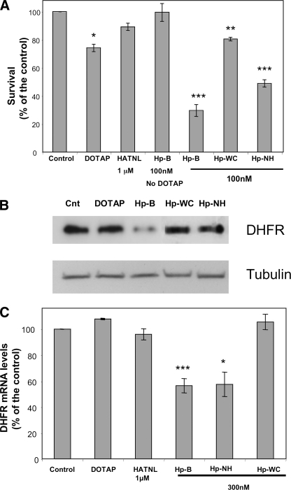
We analyzed whether polypurine hairpins (PPRHs) had the ability to knock down gene expression. These hairpins are formed by two antiparallel purine domains linked by a loop that allows the formation of Hoogsteen bonds between both domains and Watson-Crick bonds with the target polypyrimidine sequence, forming triplex structures. To set up the experimental conditions, the human dhfr gene was used as a model. The PPRHs were designed toward the template strand of DNA. The transfection of the human breast cancer cell line SKBR3 with these template hairpins against the dhfr gene produced higher than 90% of cell mortality. Template PPRHs produced a decrease in DHFR mRNA, protein, and its corresponding enzymatic activity. In addition, the activity of DHFR PPRHs was tested against breast cancer cells resistant to methotrexate, observing high cell mortality. Given the difficulty in finding long polypyrimidine stretches, we studied how to compensate for the presence of purine interruptions in the polypyrimidine target sequence. The stability of PPRH was measured, resulting in a surprisingly long half-life of about 5 days. Finally, to test the generality of usage, template PPRHs were employed against two important genes involved in cell proliferation, telomerase and survivin, producing 80 and 95% of cell death, respectively. Taken together our results show the ability of antiparallel purine hairpins to bind the template strand of double strand DNA and to decrease gene transcription. Thus, PPRHs can be considered as a new type of molecules to modulate gene expression.


Figures

FIGURE 1.
FIGURE 1.
Binding of PPRHs to polypurine/polypyrimidine target sequence-1 in the dhfr gene. A, schematic representation of the binding of the PPRHs to the dsDNA dhfr target-1 in intron 3 of the dhfr gene. B, binding of the different PPRHs to 20,000 cpm of [32P]-dhfr target-1. Dpx, duplex free target; Hp-B, hairpin HpdI3-B (blunted); Hp-T, hairpin HpdI3-T (tailed); Hp-BT, hairpin HpdI3-BT (tailed and blunted); Hp-WC, hairpin HpdI3-WC (Watson-Crick); Hp-NH, HpdI3-NH (no Hoogsteen) and Hp-Sc (scrambled). Arrows indicate the bands corresponding to the different species (duplex or triplex) in the image obtained by PhosphorImaging.
FIGURE 2.
FIGURE 2.
Cytotoxicity caused by template-PPRHs designed against dhfr target-1. A, comparison of the cytotoxic effect of HpdI3-B with that of the negative controls HpdI3-WC and HpdI3-Sc, and the no Hoogsteen variant at a concentration of 500 nm transfected with 10 μm DOTAP. The effect of the antisense HATNL-24 at 1 μm was also tested. B, study of the best PPRH:DOTAP molar ratio to attain maximum cytotoxicity. Hairpin HpdI3-B was incubated at varying concentrations in combination with a fixed concentration of 10 μm DOTAP. C, different template structures related to HpdI3-B were tested: HpdI3-C (contrary), HpdI3-T (tailed), HpdI3-BT (tailed and blunted), all at a concentration of 1 μm transfected with 10 μm DOTAP. D, determination of the optimal time for PPRH action. The medium of the cell was changed at the indicated times after transfection of 100 nm HpdI3-B with 10 μm DOTAP. For all conditions, 10,000 SKBR3 cells were plated in F-12-GHT medium and 1 week after treatment an MTT assay was performed. Data represent the mean ± S.E. of at least four experiments. *, p < 0.05; **, p < 0.01; ***, p < 0.005.
FIGURE 3.
FIGURE 3.
DHFR activity and mRNA levels upon incubation of SKBR3 cells with template-PPRHs. A, dose response of the effect of hairpin HpdI3-B on DHFR activity. 10,000 SKBR3 cells were incubated in Ham's F-12 medium lacking thymidine either with 10 μm DOTAP alone or with HpdI3-B at the indicated concentrations. HpdI3-NH and HpdI3-WC were also used at 100 nm. Two days after transfection, 6-[3H]deoxyuridine was added for 24 h to determine DHFR activity by its incorporation into DNA. Data represent the mean ± S.E. of three experiments. B, dose response of the effect of hairpin HpdI3-B on DHFR mRNA levels. 30,000 SKBR3 cells were incubated either with 10 μm DOTAP alone or HpdI3-B at the indicated concentrations. Negative controls, Hp-WC and Hp-Sc as well as Hp-NH were used at 100 nm. For comparative purposes the effect of aODN HATNL-24 was also tested at 1 μm. Three days after transfection, DHFR mRNA levels were determined by RT-real time-PCR using APRT mRNA to normalize the results. Data represent the mean ± S.E. of five experiments. *, p < 0.05; **, p < 0.01; ***, p < 0.005.
FIGURE 4.
FIGURE 4.
Effects of template-PPRHs directed against the dhfr gene in MCF7 cells resistant to 10-6m methotrexate. A, cytotoxicity. 10,000 MCF7-R cells were plated in DHFR selective medium (-GHT). Cells were incubated with hairpins HpdI3-B, HpdI3-NH, or HpdI3-WC at the indicated concentrations and 10 μm DOTAP. A week after treatment an MTT assay was performed. Data represent the mean ± S.E. of at least four experiments. B, DHFR protein levels. 30,000 MCF7-R cells were incubated with DOTAP alone (30 μm), or in combination with 300 nm of the indicated PPRHs. Three days after transfection, DHFR protein levels were determined by Western blot using tubulin to normalize the results. A representative image of DHFR and tubulin levels after PPRH treatment is shown. C, DHFR mRNA levels, 30,000 MCF7-R cells were incubated with 30 μm DOTAP alone, aODN HATNL-24, and the indicated PPRHs. Three days after transfection, DHFR mRNA levels were determined by RT-real time-PCR using the signal corresponding to APRT mRNA to normalize the results. Data represent the mean ± S.E. of at least four experiments. *, p < 0.05; **, p < 0.01; ***, p < 0.005.
FIGURE 5.
FIGURE 5.
Uptake and degradation of HpdI3-BF. 100,000 SKBR3 cells were plated in Ham's F-12 medium and incubated with 500 nm HpdI3-BF and 10 μm DOTAP. A, cells were harvested at different times and intracellular fluorescence was determined by flow cytometry. B, 24 h after transfection the medium was renewed to eliminate the remaining HpdI3-BF. Cells were harvested at different times and intracellular fluorescence was determined by flow cytometry. HpdI3-BF half-life was obtained after calculating the curve fit. Data represent the mean of three experiments.
FIGURE 6.
FIGURE 6.
Binding and associated cytotoxicity of PPRHs to target sequences 2 and 3 containing purine interruptions in the dhfr gene. A, schematic representation of the binding of the PPRHs HpdI3-misTA, -misTG and -misTT to the dsDNA dhfr target-2 bearing one adenine in the polypyrimidine stretch, and B, binding of HpdI3-misCA to dhfr target-3 bearing one guanine in the polypyrimidine sequence. Both DNA targets are located in intron 3 of the dhfr gene. A, binding of PPRHs HpdI3-misTA, -misTG, and -misTT; and B, of HpdI3-misCA to 20,000 cpm of [32P]-dhfr target-2 and [32P]-dhfr target-3, respectively, either as a duplex (Dpx) or ssDNA species. For indicates the polypurine strand of the duplex, whereas Rev represent the polypyrimidine strand. C, cytotoxicity caused by the incubation of 10,000 SKBR3 cells in DHFR selective medium (-GHT) upon transfection with the indicated PPRHs and 10 μm DOTAP. A week after treatment an MTT assay was performed. ssDNA, single-stranded DNA. Data represent the mean ± S.E. of at least four experiments. *, p < 0.05; **, p < 0.01.
FIGURE 7.
FIGURE 7.
Effect of template-PPRHs against the telomerase gene. A, schematic representation of the PPRHs used to target intron 8 in the telomerase gene. B, cytotoxicity. 10,000 SKBR3 cells were plated in Ham's F-12 medium. Cells were incubated with 10 μm DOTAP alone, HptI8-B without DOTAP, HptI8-B, HpdtI8-WC, and HptI8-Sc. A week after treatment an MTT assay was performed. C, telomerase mRNA levels. 30,000 SKBR3 cells were incubated with 10 μm DOTAP alone, HptI8-B, HpdtI8-WC, or HptI8-Sc. Three days after transfection, telomerase mRNA levels were determined by RT-real time-PCR using APRT mRNA to normalize the results. Data represent the mean ± S.E. of at least four experiments. **, p < 0.01; ***, p < 0.005.
FIGURE 8.
FIGURE 8.
Effect of template-PPRHs against the survivin gene. A, schematic representation of the PPRHs used to target exon 3 and the promoter of the survivin gene. B, cytotoxicity. 10,000 SKBR3 cells were plated in Ham's F-12 medium. Cells were incubated with either 10 μm DOTAP alone, or HpsPr-B and HpsEx3-B without DOTAP, HpsPr-B, HpsEx3-B, HpsPr-WC, or HpsPr-Sc. A week after treatment an MTT assay was performed. C, survivin mRNA levels. 30,000 SKBR3 cells were incubated with either 10 μm DOTAP alone, HpsPr-B, HpsPr-WC, or HpsPr-Sc. Three days after transfection, survivin mRNA levels were determined by RT-real time-PCR using APRT mRNA to normalize the results. Data represent the mean ± S.E. of at least four experiments. *, p < 0.05; ***, p < 0.005.

Similar articles

Cited by

References

    1. Chan, P. P., and Glazer, P. M. (1997) J. Mol. Med. 75, 267-282 - PubMed
    1. Giovannangeli, C., and Hâeláene, C. (1997) Antisense Nucleic Acid Drug Dev. 7, 413-421 - PubMed
    1. Casey, B. P., and Glazer, P. M. (2001) Prog. Nucleic Acids Res. Mol. Biol. 67, 163-192 - PubMed
    1. Felsenfeld, G., and Rich, A. (1957) Biochim. Biophys. Acta 26, 457-468 - PubMed
    1. Ryan, K., and Kool, E. T. (1998) Chem. Biol. 5, 59-67 - PubMed

Publication types

LinkOut - more resources