. 2009 Mar 15;23(6):740-54.
doi: 10.1101/gad.1739609. Epub 2009 Mar 4.

MERIT40 controls BRCA1-Rap80 complex integrity and recruitment to DNA double-strand breaks

Affiliations

MERIT40 controls BRCA1-Rap80 complex integrity and recruitment to DNA double-strand breaks

Genze Shao et al. Genes Dev. .

Abstract

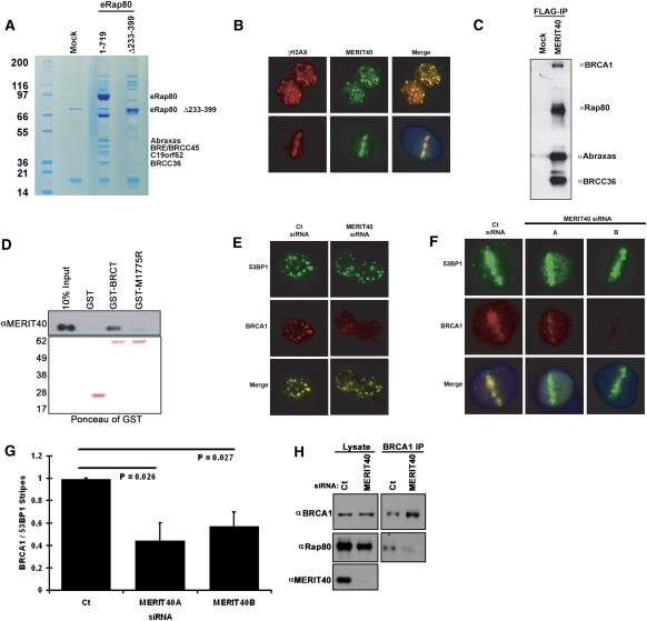
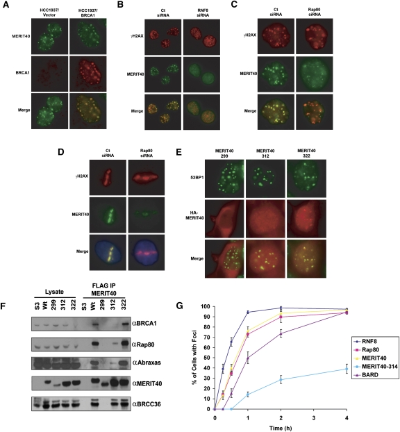
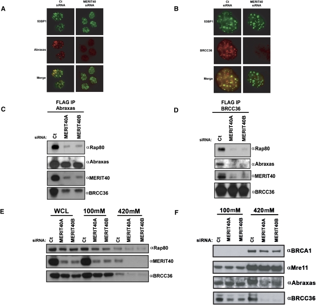
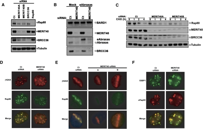
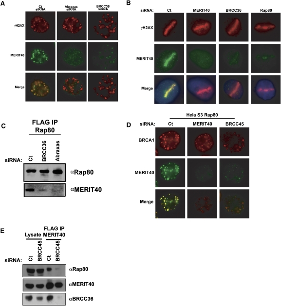
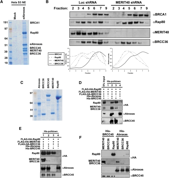
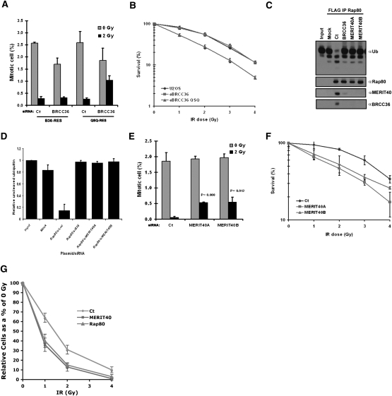
Rap80 targets the breast cancer suppressor protein BRCA1 along with Abraxas and the BRCC36 deubiquitinating enzyme (DUB) to polyubiquitin structures at DNA double-strand breaks (DSBs). These DSB targeting events are essential for BRCA1-dependent DNA damage response-induced checkpoint and repair functions. Here, we identify MERIT40 (Mediator of Rap80 Interactions and Targeting 40 kD)/(C19orf62) as a Rap80-associated protein that is essential for BRCA1-Rap80 complex protein interactions, stability, and DSB targeting. Moreover, MERIT40 is required for Rap80-associated lysine(63)-ubiquitin DUB activity, a critical component of BRCA1-Rap80 G2 checkpoint and viability responses to ionizing radiation. Thus, MERIT40 represents a novel factor that links BRCA1-Rap80 complex integrity, DSB recognition, and ubiquitin chain hydrolytic activities to the DNA damage response. These findings provide new molecular insights into how BRCA1 associates with independently assembled core protein complexes to maintain genome integrity.


Figures

Figure 1.
Figure 1.
Identification of MERIT40 as a novel BRCA1–Rap80 complex constituent that is required for BRCA1 DSB localization. (A) Coomassie-stained gel representing eRap80 (1–719 and Δ233–399) complexes after consecutive Flag and HA affinity-purification steps from HeLa S3 nuclear extracts. The identities of both eRap80 species and the four Coomassie-stained bands that are uniquely present in the eRap80 wild-type complex are indicated as Abraxas, BRE/BRCC45, BRCC36, and C19orf62. (B) IF for endogenous MERIT40 in HeLa cells at 4 h following IR (top panel) and 30 min after laser microirradiation (bottom panel), demonstrating MERIT40 colocalization with γH2AX at DSBs. (C) Immunoblot (IB) of the eMERIT40 complex after Flag IP from HeLa S3 cells. BRCC36, Abraxas, Rap80, and BRCA1 were detected as MERIT40-associated proteins. (D) MERIT40 interacts with the BRCA1 BRCT domain, but not a clinical BRCT domain mutant. Nuclear extracts of HeLa S3 cells expressing eMERIT40 were incubated with recombinant GST, GST-BRCT, or GST-M1775R proteins bound to GST beads. (Top) IB with anti-HA antibody was performed to detect eMERIT40. (Bottom) The Ponceau-stained membrane is displayed showing the amount of each GST fusion protein. (E,F) MERIT40 is required for BRCA1 DSB localization. IF was performed for BRCA1 following Ct or MERIT40 siRNA treatment in IR-treated (E) and laser-microirradiated (F) HeLa cells. MERIT40 siRNA A and B target different sequences in the coding region of the MERIT40 mRNA. (G) Quantification of BRCA1 stripes as a percentage of 53BP1 stripes displayed graphically from F. At least 200 cells were counted in triplicate for the analysis. Error bars represent standard deviation (SD) and P values were calculated by the Student's t-test. (H) MERIT40 is required for BRCA1 association with Rap80. BRCA1 IP was performed in HeLa S3 cells following transfection with MERIT40 or Ct siRNA. IB was performed on the BRCA1 IP eluates as indicated.
Figure 2.
Figure 2.
MERIT40 DSB localization depends on the RNF8 E3 ligase and interaction with Rap80. (A) IF for MERIT40 and BRCA1 in irradiated HCC1937 cells reconstituted with vector (left panels) or wild-type BRCA1 (right panels), demonstrating MERIT40 localization to IRIF occurs independently of wild-type BRCA1 status. (B) IF for MERIT40 and γH2AX in IR-treated HeLa cells following Ct or RNF8 siRNA treatment, demonstrating dependence on RNF8 for MERIT40 IRIF. (C,D) IF for MERIT40 and γH2AX in irradiated (C) and laser-microirradiated (D) HeLa cells following Ct or Rap80 siRNA treatment demonstrates reduced MERIT40 DSB localization in Rap80-depleted cells. (E) IF detection by α-HA antibodies of ectopically expressed forms of Flag-HA-MERIT40 following IR. (Left panel) MERIT40-299 (residues 1–299) is cytoplasmic. (Middle panel) MERIT40-312 (residues 1–312) is nuclear and weakly colocalizes with 53BP1. (Right panel) MERIT40-322 (residues 1–322) strongly colocalizes with 53BP1, akin to full-length MERIT40 (residues 1–329). (F) IB of lysates and Flag IP elutions from HeLa S3 cells expressing epitope-tagged MERIT40 truncations (299, 312, 322, and wild type). MERIT40-299 loses all interactions whereas MERIT40-312 demonstrates reduced interaction with Rap80, Abraxas, and BRCC36. MERIT40-322 interacts with Rap80, Abraxas, and BRCC36. (G) Kinetics of Flag-HA-tagged RNF8, Rap80, MERIT40, MERIT40-314, and BARD IRIF formation in HeLa S3 cells following 10 Gy IR, showing MERIT40 localization occurs more slowly than RNF8 but faster than BARD IRIF. At least 200 cells were counted in triplicate for the analysis. Error bars represent SD. Representative cells for each time point are shown in Supplemental Figure S5.
Figure 3.
Figure 3.
MERIT40 is required for DSB targeting and chromatin association of Abraxas and BRCC36. (A,B) MERIT40 knockdown reduces Abraxas (A) and BRCC36 (B) DSB localization. IF was performed for Abraxas and BRCC36 in cells transfected with Ct or MERIT40 siRNA at 4 h after 10 Gy IR. Images are representative of >200 cells counted per condition in three independent experiments. (C,D) HeLa S3 cells expressing eAbraxas (C) and eBRCC36 (D) were immunoprecipitated with anti-Flag-M2 Agarose beads at 72 h after transfection with Ct or either MERIT40A- or MERIT40B-targeted siRNAs. IB of Flag elutions was performed as indicated, demonstrating that both Abraxas and BRCC36 require MERIT40 for interaction with Rap80. (E,F) MERIT40 depletion results in reduced levels of Rap80 and BRCC36 (E) and Abraxas (F) in the high-salt extractable fractions, as shown by IB of sequential 100 mM and 420 mM NaCl-containing lysates.
Figure 4.
Figure 4.
MERIT40 mediates the stability of the BRCA1–RAP80 complex at DSBs. (A) MERIT40 depletion results in decreased Rap80 and BRCC36 protein levels. IB was performed on HeLa cell lysates at 72 h following siRNA transfection as indicated. (B) MERIT40 is required for endogenous Abraxas stability (left) and ectopic Abraxas in an eAbraxas cell line (right). IB was performed for Abraxas in nontransduced (mock) or eAbraxas-expressing HeLa cells at 72 h after Ct or MERIT40 siRNA. Note that eAbraxas expression causes reduced levels of endogenous Abraxas protein. (C) MERIT40 depletion reduces Rap80 and BRCC36 protein levels and half-life. HeLa cells were transfected with Ct or MERIT40 siRNAs. Untreated (0) or treated cells were lysed at the indicated time following the addition of CHX (60 μg/mL). IB was performed as indicated. (D,E) MERIT40 knockdown reduces Rap80 DSB localization. IF was performed for Rap80 following Ct or MERIT40 siRNA treatment at IR-induced (D) or laser-induced (E) DSBs as indicated. Images are representative of >200 cells for each condition in three independent experiments. (E) Quantification of Rap80 colocalization with γH2AX at stripes is shown in Supplemental Figure S8A. (F) HeLa S3 cells containing stably expressed eRap80 were transfected with Ct- or MERIT40-targeted siRNA and subsequently examined for IRIF with α-HA and α-53BP1 antibodies. eRap80 is still able to localize to IRIF following MERIT40 depletion.
Figure 5.
Figure 5.
Interdependence of the BRCA1–Rap80 complex for DSB localization and protein interactions. (A) IF for MERIT40 and γH2AX at 4 h after 10 Gy IR in HeLa cells following Ct (left panel), Abraxas (middle panel), or BRCC36 (right panel) siRNA treatment demonstrates reduced MERIT40 DSB localization. Results are quantified and displayed graphically in Supplemental Figure S4B. (B) IF for MERIT40 and γH2AX in laser-irradiated HeLa cells following Ct, MERIT40, BRCC36, or Rap80 siRNA treatment demonstrates reduced MERIT40 DSB localization. Images are representative of >200 cells for each condition in three independent experiments. (C) Requirement for BRCC36 and Abraxas in Rap80 interactions with MERIT40. Flag IP was performed on HeLa S3 cells expressing ectopic Rap80 at 72 h after transfection with Ct-, BRCC36-, or Abraxas-targeted siRNAs. IB of Flag elutions was performed as indicated, demonstrating that both Abraxas and BRCC36 are required for Rap80 interaction with MERIT40. (D) IF for BRCA1 and MERIT40 following Ct (left panel), MERIT40 (middle panel), or BRCC45 (right panel) siRNA treatment in HeLa S3 cells stably expressing ectopic Flag-HA tagged Rap80. Depletion of MERIT40 or BRCC45 reduces BRCA1 IRIF. Images are representative of >200 cells for each condition in three independent experiments. (E) Requirement for BRCC45 in MERIT40 interaction with Rap80 and BRCC36. Flag IP was performed on HeLa S3 cells expressing ectopic MERIT40 at 72 h after transfection with Ct- or BRCC45-targeted siRNAs. IB of Flag elutions was performed as indicated, demonstrating that BRCC45 is required for MERIT40 interaction with Rap80 and BRCC36.
Figure 6.
Figure 6.
Biochemical reconstitution of the Rap80 complex in vitro. (A) Coomassie-stained gel representing eAbraxas complexes after consecutive Flag and HA affinity-purification steps from HeLa S3 nuclear extracts (NE). The predominant interacting proteins are readily observed in strong Coomassie-stained bands for Rap80, eAbraxas, BRCC45, MERIT40, and BRCC36. BRCA1 was detected substoichiometrically as an Abraxas-associated protein. (B) HeLa S3 nuclear extracts derived from cells expressing shRNA to either luciferase (luc) or MERIT40 were centrifuged through a 5%–22.5% sucrose gradient, and individual fractions were probed by IB as indicated. MERIT40 depletion shifts peak protein levels of Rap80 and BRCC36 to lower fractions. Quantification of bands was performed using ImageJ software from the NIH. (C) Coomassie-stained gel representing purified proteins used for in vitro binding assays. Recombinant Abraxas and BRCC45 are His-NusA- and His-ZZ-tagged, respectively; were bacterially derived; and are estimated to be ∼90% pure. Recombinant BRCC36, MERIT40, and Rap80 derived from baculovirus-infected Sf9 cells are Flag-HA-tagged and are estimated to be ∼95% pure. (D,E) IB of in vitro binding assays was performed as indicated. BRCC36, MERIT40, and Rap80 were baculovirally expressed and mixed in equal molar concentrations with both Abraxas and BRCC45 or combinations thereof. HisPur Cobalt resin was used for coprecipitation of recombinant proteins. (F) IB of in vitro binding assays was performed as indicated. His-tagged BRCC45 or His-tagged Abraxas were mixed in equal molar concentration with Rap80, MERIT40, or BRCC36 individually and coprecipitated using HisPur Cobalt resin.
Figure 7.
Figure 7.
MERIT40 and BRCC36 DUB contributions to the DDR. (A) Knockdown of endogenous BRCC36 in U2OS cells that ectopically express siRNA-resistant forms of either B36 wild type or B36 QSQ reveals that BRCC36 DUB activity is required for the G2 checkpoint. Mitotic cells were detected by FACS with an antibody against phosphohistone H3. Error bars represent SD. (B) BRCC36 DUB deficiency increases IR sensitivity as measured by colony formation in U2OS cells that express eBRCC36 wild type or eBRCC36 QSQ. Each group was plated at low density and exposed to escalating doses of IR as indicated. Cells were maintained in culture for 12 d following IR and subsequently Giemsa-stained. Colonies were counted and normalized as a percentage of colonies formed at 0 Gy. Each point represents the average of three independent experiments. Error bars indicate SD. (C) Knockdown with either BRCC36 or MERIT40 siRNA reduces Rap80-associated K63-Ub DUB activity. eRap80 complexes were purified from nuclear extracts of HeLa S3 cells at 48 h after transfection with the indicated siRNAs. Mock represents Flag IP from HeLa S3 cells that do not express ectopic protein. Flag-purified complexes were incubated with hexa-K63–Ub, and the products were detected by IB with antibody to Ub. The figure is representative of three independent experiments. (D) Quantification of C using ImageJ software from the NIH. Relative uncleaved ubiquitin represents the relative intensity of the uppermost K63–Ub band in the IB normalized to input. Error bars represent SD. (E) MERIT40 is required for the G2 checkpoint. The G2 checkpoint assay was performed as in A following Ct or MERIT40 siRNAs as indicated. The percentage of mitotic cells is graphically displayed. Error bars represent SD and P values were calculated by the Student's t-test for each MERIT40 siRNA compared with Ct siRNA. (F) Knockdown of endogenous MERIT40 in HeLa cells reveals increased IR sensitivity. Colony formation assay was performed as in B following Ct or MERIT40 siRNAs. Cells were treated with the indicated doses of IR at 48 h after siRNA transfection. Error bars represent SD. (G) MERIT40 and Rap80 contribute to IR resistance in BRCA1 mutated cells. HCC1937 cells were exposed to escalating doses of IR 48 h after transfection with the indicated siRNAs. Relative cell number at each dose of IR is displayed graphically. Error bars represent SD.

Similar articles

Cited by

References

    1. Ambroggio X.I., Rees D.C., Deshaies R.J. JAMM: A metalloprotease-like zinc site in the proteasome and signalosome. PLoS. 2004;2:e2. doi: 10.1371/journal.pbio.0020002. - DOI - PMC - PubMed
    1. Bassermann F., Frescas D., Guardavaccaro D., Busino L., Peschiaroli A., Pagano M. The Cdc14B–Cdh1–Plk1 axis controls the G2 DNA-damage-response checkpoint. Cell. 2008;134:256–267. - PMC - PubMed
    1. Bekker-Jensen S., Lukas C., Kitagawa R., Melander F., Kastan M.B., Bartek J., Lukas J. Spatial organization of the mammalian genome surveillance machinery in response to DNA strand breaks. J. Cell Biol. 2006;173:195–206. - PMC - PubMed
    1. Bridge W.L., Vandenberg C.J., Franklin R.J., Hiom K. The BRIP1 helicase functions independently of BRCA1 in the Fanconi anemia pathway for DNA crosslink repair. Nat. Genet. 2005;37:953–957. - PubMed
    1. Celeste A., Fernandez-Capetillo O., Kruhlak M.J., Pilch D.R., Staudt D.W., Lee A., Bonner R.F., Bonner W.M., Nussenzweig A. Histone H2AX phosphorylation is dispensable for the initial recognition of DNA breaks. Nat. Cell Biol. 2003;5:675–679. - PubMed

Publication types