. 2009 Mar 24;106(12):4894-9.
doi: 10.1073/pnas.0811507106. Epub 2009 Mar 4.

Phasic excitation of dopamine neurons in ventral VTA by noxious stimuli

Affiliations

Phasic excitation of dopamine neurons in ventral VTA by noxious stimuli

Frédéric Brischoux et al. Proc Natl Acad Sci U S A. .

Abstract

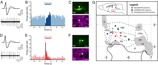
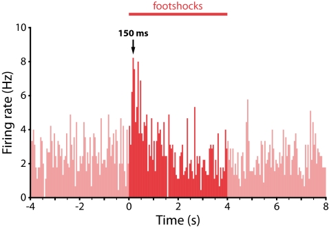
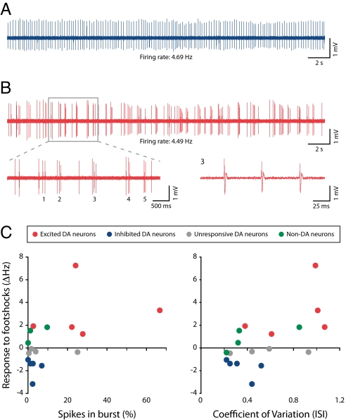
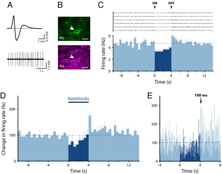
Midbrain dopamine neurons play central roles in reward processing. It is widely assumed that all dopamine neurons encode the same information. Some evidence, however, suggests functional differences between subgroups of dopamine neurons, particularly with respect to processing nonrewarding, aversive stimuli. To directly test this possibility, we recorded from and juxtacellularly labeled individual ventral tegmental area (VTA) dopamine neurons in anesthetized rats so that we could link precise anatomical position and neurochemical identity with coding for noxious stimuli. Here, we show that dopamine neurons in the dorsal VTA are inhibited by noxious footshocks, consistent with their role in reward processing. In contrast, we find that dopamine neurons in the ventral VTA are phasically excited by footshocks. This observation can explain a number of previously confusing findings that suggested a role for dopamine in processing both rewarding and aversive events. Taken together, our results indicate that there are 2 functionally and anatomically distinct VTA dopamine systems.


Conflict of interest statement

The authors declare no conflict of interest.

Figures

Fig. 1.
Fig. 1.
Dorsal VTA dopamine neurons are inhibited by noxious stimuli, whereas ventral VTA dopamine neurons are excited. (A) Averaged extracellular waveform and baseline firing activity from a recorded neuron. (B and C) This neuron (B) showed an inhibitory response to footshocks (peristimulus time histogram averaged across 6 footshocks; mean + SEM; 500-ms bins) and was (C) immunohistochemically identified as dopaminergic (Nb indicates Neurobiotin). (D–F) In contrast, a second neuron with a similar averaged extracellular waveform and baseline firing rate (D) showed an excitatory response to footshocks (E), but was also immunohistochemically identified as dopaminergic (F). (Scale bars: 20 μm.) (G) A parasagittal schematic view of the VTA (lateral, 0.6 mm) showing the distribution of individual dopamine neurons and their responses to footshocks and showing a clear anatomical segregation of functional subgroups (horizontal numbers are distance from bregma in millimeters; vertical numbers are depth in millimeters). fr indicates fasciculus retroflexus; IP, interpeduncular nucleus; ml, medial lemniscus; mp, mammillary peduncle; PBP, parabrachial pigmented nucleus; PFR, parafasciculus retroflexus area; PIF, parainterfascicular nucleus; PN, paranigral nucleus; rs, rubrospinal tract; tth, trigeminothalamic tract; and VTAc, ventral tegmental area caudal.
Fig. 2.
Fig. 2.
Footshock-evoked excitations in ventral VTA dopamine neurons have a rapid onset, similar to that seen for reward-related excitations in dopamine neurons in previous studies. Population peristimulus time histogram for the 5 identified dopamine neurons that exhibited an excitatory response to footshocks (50-ms bins).
Fig. 3.
Fig. 3.
Dopamine neurons that are inhibited by the noxious stimulus show tonic, low-bursting, single-spike activity, whereas dopamine neurons that are excited by the noxious stimulus show high levels of bursting. (A) Tonic firing activity in an identified dopamine neuron that was inhibited by the footshocks. (B) Bursting activity in an identified dopamine neuron that was excited by the footshocks (Upper). (Lower) An expanded view of 5 bursts (Left) and the third burst (Right). (C) Scatter plots showing change in response to footshock as a function of the percentage of spikes in burst (Left) or CV of the ISI (Right). Dopamine neurons that were excited by the footshocks had higher levels of bursting and higher ISI CVs than either dopamine neurons that were inhibited by the footshocks or nondopamine neurons [1 nondopamine neuron could not be analyzed for bursts because its high-baseline firing rate (9.14 Hz) meant that its mean ISI fell within the burst criteria].
Fig. 4.
Fig. 4.
Many dopamine neurons that are inhibited by (or unresponsive to) the footshocks show a significant excitation at the termination of the stimulus. (A) Averaged extracellular waveform and baseline firing activity from a recorded neuron. (B) The same neuron was immunohistochemically identified as dopaminergic (Nb indicates Neurobiotin). (Scale bars: 20 μm.) (C) A cumulative raster plot of this neuron (Upper) and the resulting peristimulus time histogram (Lower) averaged across 6 footshocks (mean + SEM; 500-ms bins) showed an inhibitory response following the onset (ON) of the stimulation and an excitation following its offset (OFF). Black dashed line indicates mean baseline firing rate. (D) Population peristimulus time histograms (500-ms bins; mean + SEM) for the 5 identified dopamine neurons that exhibited an excitation at the offset of the footshocks. (E) Higher-resolution population peristimulus time histograms (50-ms bins; mean) showing latency of the peak response at the offset of footshocks.

Comment in

  • The dopamine puzzle.
    Schmidt R, Morris G, Hagen EH, Sullivan RJ, Hammerstein P, Kempter R. Schmidt R, et al. Proc Natl Acad Sci U S A. 2009 Jul 7;106(27):E75. doi: 10.1073/pnas.0905153106. Epub 2009 Jun 19. Proc Natl Acad Sci U S A. 2009. PMID: 19549815 Free PMC article. No abstract available.
  • Duality of salience in dopamine neurons.
    Root DH, Barker DJ, Ma S. Root DH, et al. Proc Natl Acad Sci U S A. 2009 Aug 4;106(31):E84. doi: 10.1073/pnas.0906641106. Epub 2009 Jul 23. Proc Natl Acad Sci U S A. 2009. PMID: 19628687 Free PMC article. No abstract available.

References

    1. Schultz W. Predictive reward signal of dopamine neurons. J Neurophysiol. 1998;80:1–27. - PubMed
    1. Wise RA. Dopamine, learning and motivation. Nat Rev Neurosci. 2004;5:483–494. - PubMed
    1. Ford CP, Mark GP, Williams JT. Properties and opioid inhibition of mesolimbic dopamine neurons vary according to target location. J Neurosci. 2006;26:2788–2797. - PMC - PubMed
    1. Lammel S, et al. Unique properties of mesoprefrontal neurons within a dual mesocorticolimbic dopamine system. Neuron. 2008;57:760–773. - PubMed
    1. Margolis EB, Mitchell JM, Ishikawa J, Hjelmstad GO, Fields HL. Midbrain dopamine neurons: Projection target determines action potential duration and dopamine D2 receptor inhibition. J Neurosci. 2008;28:8908–8913. - PMC - PubMed

Publication types