. 2009 Mar 4;29(9):2754-61.
doi: 10.1523/JNEUROSCI.4413-08.2009.

Exercise reduces GABA synaptic input onto nucleus tractus solitarii baroreceptor second-order neurons via NK1 receptor internalization in spontaneously hypertensive rats

Affiliations

Exercise reduces GABA synaptic input onto nucleus tractus solitarii baroreceptor second-order neurons via NK1 receptor internalization in spontaneously hypertensive rats

Chao-Yin Chen et al. J Neurosci. .

Abstract

A single bout of mild to moderate exercise can lead to a postexercise decrease in blood pressure in hypertensive subjects, namely postexercise hypotension (PEH). The full expression of PEH requires a functioning baroreflex, hypertension, and activation of muscle afferents (exercise), suggesting that interactions in the neural networks regulating exercise and blood pressure result in this fall in blood pressure. The nucleus tractus solitarii (NTS) is the first brain site that receives inputs from nerves carrying blood pressure and muscle activity information, making it an ideal site for integrating cardiovascular responses to exercise. During exercise, muscle afferents excite NTS GABA neurons via substance P and microinjection of a substance P-neurokinin 1 receptor (NK1-R) antagonist into the NTS attenuates PEH. The data suggest that an interaction between the substance P NK1-R and GABAergic transmission in the NTS may contribute to PEH. We performed voltage clamping on NTS baroreceptor second-order neurons in spontaneously hypertensive rats (SHRs). All animals were killed within 30 min and the patch-clamp recordings were performed 2-8 h after the sham/exercise protocol. The data showed that a single bout of exercise reduces (1) the frequency but not the amplitude of GABA spontaneous IPSCs (sIPSCs), (2) endogenous substance P influence on sIPSC frequency, and (3) sIPSC frequency response to exogenous application of substance P. Furthermore, immunofluorescence labeling in NTS show an increased substance P NK1-R internalization on GABA neurons. The data suggest that exercise-induced NK1-R internalization results in a reduced intrinsic inhibitory input to the neurons in the baroreflex pathway.


Figures

Figure 1.
Figure 1.
A, An NTS baroreceptor second-order neuron. a, The neuron viewed with infrared differential interference contrast (IR-DIC). b, The labeled aortic depressor nerve boutons viewed with fluorescence filter set. c, Overlay of the IR-DIC and fluorescence images. d, Neuron with patch electrode in whole-cell configuration. Scale bar, 10 μm. B, Photomicrograph shows the position of the recording electrode. TS, Tractus solitarius. Scale bar, 200 μm.
Figure 2.
Figure 2.
A, Example traces of sIPSC recorded from one Sham and one PEH SHR. B, Group data of cumulative probability of sIPSC interevent interval (left) and amplitude (right). PEH was associated with a rightward shift in the interevent interval and reduced mean frequency. There was no change in the sIPSC amplitude after exercise.
Figure 3.
Figure 3.
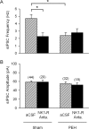
A, Group data of sIPSC frequency. Incubation with the NK1-R antagonist (SR140,333, 1 μm) significantly reduced the sIPSC frequency in Sham but not in PEH SHRs. B, Group data of sIPSC amplitude. In the same neurons, NK1-R antagonist had no effect on sIPSC amplitude in both groups. Numbers in parentheses indicate number of neurons. *p < 0.05, Fisher's LSD test.
Figure 4.
Figure 4.
A, Example traces of sIPSCs recorded before (Control), during substance P perfusion, and during washout period from one Sham and one PEH SHR. B, Group data of substance P-induced increase in sIPSC frequency. Substance P concentration-dependently increased sIPSC frequency in baroreceptor second-order neurons. The neurons recorded from PEH rats had a small response to application of substance P. Numbers in parentheses indicate number of neurons. *p < 0.05, two-way ANOVA.
Figure 5.
Figure 5.
A, Example traces of mIPSC recorded from one Sham and one PEH SHR. B, Group data showing no difference in cumulative probability of interevent interval (left) and amplitude (right).
Figure 6.
Figure 6.
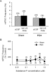
A, Group data of mIPSC frequency. Incubation with the NK1-R antagonist (NK1-R Anta.) had no effect on the mIPSC frequency in both Sham and PEH groups. B, Group data of effects of substance P application on mIPSC frequency. Substance P had no consistent effect on mIPSC frequency in baroreceptor second-order neurons. Numbers in parentheses indicate number of neurons.
Figure 7.
Figure 7.
A, Example images from one Sham (A1) and one PEH (A2) rat. a, NK1-R staining (red color). b, GAD67 staining (green color). c, SYTOX staining (blue color). d, Overlap of a, b, and c. B, Fluorescent intensity plot across the white lines in A1d and A2d. PEH was associated with a higher degree of NK1-R internalization on NTS GABA neurons.
Figure 8.
Figure 8.
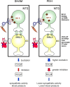
A simplified schematic of the proposed mechanism contributing to PEH in hypertensive subjects. During exercise, muscle afferent releases substance P to activate the NTS GABA interneurons to reset baroreflex to a higher blood pressure. Consequently, activation of NK1-R during exercise triggers the receptors to undergo internalization that could have a prolonged effect on baroreflex regulation after exercise. Our immunofluorescence data demonstrate that the NK1-R on the NTS GABA interneurons had a higher degree of internalization after a single bout of exercise in SHRs. The NK1-R internalization on GABA interneurons after exercise could contribute to disinhibition of the neurons in the baroreflex pathway. Our whole-cell patch recordings from the baroreceptor second-order neurons in NTS demonstrate that a single bout of exercise resulted in a reduced GABA inhibition. Disinhibition of the NTS neurons in the baroreflex pathway could translate to a higher excitatory output from NTS to CVLM and a greater inhibition to the RVLM resulting in a lower sympathetic activity and hence a lower blood pressure after exercise in the hypertensive subjects.

References

    1. Bonham AC, Chen CY. Glutamatergic neural transmission in the nucleus tractus solitarius: N-methyl-d-aspartate receptors. Clin Exp Pharmacol Physiol. 2002;29:497–502. - PubMed
    1. Boone JB, Jr, Corry JM. Proenkephalin gene expression in the brainstem regulates post-exercise hypotension. Brain Res Mol Brain Res. 1996;42:31–38. - PubMed
    1. Boscan P, Paton JF. Excitatory convergence of periaqueductal gray and somatic afferents in the solitary tract nucleus: role for neurokinin 1 receptors. Am J Physiol Regul Integr Comp Physiol. 2005;288:R262–R269. - PubMed
    1. Catelli JM, Sved AF. Enhanced pressor response to GABA in the nucleus tractus solitarii of the spontaneously hypertensive rat. Eur J Pharmacol. 1988;151:243–248. - PubMed
    1. Chandler MP, DiCarlo SE. Sinoaortic denervation prevents postexercise reductions in arterial pressure and cardiac sympathetic tonus. Am J Physiol. 1997;273:H2738–H2745. - PubMed

Publication types

MeSH terms

LinkOut - more resources