. 2009 Mar 4;29(9):2984-96.
doi: 10.1523/JNEUROSCI.3624-08.2009.

DSCAM deficiency causes loss of pre-inspiratory neuron synchroneity and perinatal death

Affiliations

DSCAM deficiency causes loss of pre-inspiratory neuron synchroneity and perinatal death

Kenji Amano et al. J Neurosci. .

Abstract

Down syndrome cell adhesion molecule (DSCAM) is a neural adhesion molecule that plays diverse roles in neural development. We disrupted the Dscam locus in mice and found that the null mutants (Dscam(-/-)) died within 24 h after birth. Whole-body plethysmography showed irregular respiration and lower ventilatory response to hypercapnia in the null mutants. Furthermore, a medulla-spinal cord preparation of Dscam(-/-) mice showed that the C4 ventral root activity, which drives diaphragm contraction for inspiration, had an irregular rhythm with frequent apneas. Optical imaging of the preparation using voltage-sensitive dye revealed that the pre-inspiratory neurons located in the rostral ventrolateral medulla and belonging to the rhythm generator for respiration, lost their synchroneity in Dscam(-/-) mice. Dscam(+/-) mice, which survived to adulthood without any overt abnormalities, also showed irregular respiration but milder than Dscam(-/-) mice. These results suggest that DSCAM plays a critical role in central respiratory regulation in a dosage-dependent manner.


Figures

Figure 1.
Figure 1.
Targeted disruption of Dscam in mice. A, Strategy for generating the targeted Dscam allele. Schematic representation of wild-type Dscam locus including exon 1 (Ex. 1, solid box), targeting construct using the cre–loxP system, initial homologously recombined flox–neo allele, and targeted Dscam knock-out allele. To obtain the exon 1-deleted knock-out allele, heterozygous mice with a flox–neo allele were mated with the CAG–cre mice. Four probes (P1, P2, P3, and P4) used for Southern blot analyses are indicated. Ac, AccI; B, BamHI; EI, EcoRI; EV, EcoRV; Hc, HincII; Hd, HindIII; M, MluI; Sp, SpeI. B, Southern blot analysis of SpeI- or EcoRV-digested genomic DNA from wild-type (+/+) or flox–neo (neo/+) mice revealed 9.8 or 4.8 kb fragments in homologous recombinant flox–neo mice; HincII/MluI- or AccI-digested genomic DNA from wild-type (+/+) or heterozygous (+/−) mice revealed 8.0 or 7.7 kb fragments in exon 1-deleted heterozygous mice. C, Northern blot analysis of RNA extracted from the brains of wild-type, Dscam+/−, and Dscam−/− mice at P0 using four parts of Dscam cDNA [Ex. 1, exon 1 (nucleotides 1–496); EC, extracellular (nucleotides 2299–2626); TM, transmembrane (nucleotides 5210–5410); CP, cytoplasmic (nucleotides 5385–5637)] as probes. All four probes detected the 8.7 kb Dscam transcript in the wild-type and Dscam+/− mice. Note that the targeted transcripts disappeared completely in Dscam−/− mice. The exon 1 probe detected a nonspecific band of 1.0 kb in addition to the 8.7 kb Dscam transcript. β-Actin probe was used as a control.
Figure 2.
Figure 2.
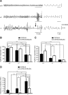
Dosage-dependent in vivo respiratory abnormalities in Dscam-deficient mice. Spontaneous respiration activities of newborn mice were measured by plethysmography. A, Recordings correspond to spontaneous respiration in wild-type, Dscam+/−, and Dscam−/− littermates, respectively (C57BL/6 background; for C57BL/6–BALB/c mixed background, see Fig. 8A). Horizontal bar under each respiration activity represents a period of 10 s (left side) or 1 s (right side), respectively. Vertical bar at the right side of respiration activity represents a volume of 0.10 mmH2O. Dscam−/− mice show irregular patterns, and Dscam+/− also show similar but less severe respiratory abnormalities. In addition, continuous lack of respiration activity representing apnea was often observed in Dscam−/− mice (arrow). B, C, Ratio of rhythmic respirations was quantified as percentage (rhythmic vs irrhythmic) or averaged time periods of continuous rhythmic respiration during quiet breathing without moving. The ratio (percentage) of rhythmic respiration (B) or averaged continuous period of rhythmic respiration (C) on C57BL/6 (filled bar) or C57BL/6–BALB/c mixed background (open bar) were calculated in wild-type (n = 10 or n = 8), Dscam+/− (n = 14 or n = 9), and Dscam−/− genotypes (n = 9 or n = 7), respectively. B, The ratio of rhythmic respiration in Dscam−/− mice was significantly lower than that in wild-type or Dscam+/− mice on both C57BL/6 and C57BL/6–BALB/c mixed backgrounds. In Dscam wild-type genotype, the ratio of rhythmic respiration on C57BL/6–BALB/c mixed background was close to but still statistically higher than that on C57BL/6 background. C, The averaged time period of continuous rhythmic respiration in Dscam−/− mice was significantly shorter than that in wild-type mice on both C57BL/6 and C57BL/6–BALB/c mixed backgrounds. The averaged period of rhythmic respiration in Dscam+/− mice was also significantly shorter than that in wild-type mice on C57BL/6 background. Note that the period of rhythmic respiration in Dscam+/− mice on C57BL/6–BALB/c mixed background was significantly longer than that in Dscam+/− mice on C57BL/6 background. The period of rhythmic respiration in Dscam wild type on C57BL/6–BALB/c mixed background was longer than that in Dscam+/− mice on C57BL/6 background but statistically not significant. D, Frequency of apnea on C57BL/6 (filled bar) or C57BL/6-BALB/c mixed background (open bar) was quantified as the number of apnea that continued without respiration for >1 s in wild-type (n = 10 or n = 9), Dscam+/− (n = 14 or n = 9), and Dscam−/− genotypes (n = 9 or n = 7), respectively. The frequency of apnea in Dscam−/− mice was significantly higher than that in wild-type or Dscam+/− mice on both C57BL/6 and C57BL/6–BALB/c mixed backgrounds. Note that the frequency of apnea in Dscam−/− mice on C57BL/6 background was significantly higher than that in Dscam−/− mice on C57BL/6–BALB/c mixed background. Statistics on Dscam genotypes with three entities were calculated by Tukey–Kramer test. Those on strains with two entities (C57BL/6 and C57BL/6–BALB/c mixed backgrounds) were calculated by Student's t test. *p < 0.05, **p < 0.01, and ***p < 0.001. Error bars represent SEM.
Figure 3.
Figure 3.
Dosage-dependent abnormalities of in vitro inspiratory activity in Dscam-deficient mice. C4 ventral root inspiratory activities in medulla–spinal cord preparations of newborn mice were measured. A, Recordings correspond to C4 inspiratory activity in wild-type, Dscam+/−, and Dscam−/− littermates, respectively (C57BL/6 background; for C57BL/6–BALB/c mixed background, see Fig. 8B). Horizontal bar under each C4 activity represents a period of 1 min. Vertical bar at the right side of C4 activity represents an amplitude of 0.20 mV. The activities for Dscam−/− show irregular patterns. Lack of large C4 inspiratory activities representing apnea was often observed in Dscam−/− mice (arrows). The activities of Dscam+/− mice were also irregular but less severe than that of Dscam−/− mice. B, Nerve activity (bursts per minute) on C57BL/6 (filled bar) or C57BL/6–BALB/c mixed background (open bar) was calculated in wild-type (n = 7 or n = 5), Dscam+/− (n = 8 or n = 5), and Dscam−/− genotypes (n = 11 or n = 5), respectively. The frequency of C4 inspiratory activity in Dscam−/− mice was significantly less than that in wild-type or Dscam+/− mice on C57BL/6 background. The C4 activity in Dscam+/− and Dscam−/− mice on C57BL/6–BALB/c mixed background was less frequent than that in wild-type mice but statistically not significant. C, Frequency of apnea, which was defined as lack of large C4 inspiratory activities for >15 s, on C57BL/6 (filled bar) or C57BL/6–BALB/c mixed background (open bar), was quantified in wild-type (n = 5 or n = 3), Dscam+/− (n = 5 or n = 3), and Dscam−/− genotypes (n = 5 or n = 5), respectively. The frequency of apnea in Dscam−/− mice was significantly higher than that in wild type on both C57BL/6 and C57BL/6–BALB/c mixed backgrounds. That of Dscam−/− was also significantly higher than that in Dscam+/− on C57BL/6 background. Although the frequency of apnea in Dscam−/− was lower than that in Dscam+/− on C57BL/6–BALB/c mixed background, the difference was not statistically significant. Note that the frequency of apnea in Dscam−/− on C57BL/6 background was significantly higher than that on C57BL/6–BALB/c mixed background. No apnea was found in wild-type mice on both C57BL/6 and C57BL/6–BALB/c mixed backgrounds. Statistics on Dscam genotypes with three entities were calculated by Tukey–Kramer test. Those on strains with two entities (C57BL/6 and C57BL/6–BALB/c mixed backgrounds) were calculated by Student's t test. *p < 0.05, **p < 0.01, and ***p < 0.001. Error bars represent SEM.
Figure 4.
Figure 4.
Abnormal ventilatory response to hypercapnia in Dscam-deficient mice. Spontaneous respiration activity in air or in a hypercapnia condition (8% CO2) was measured by plethysmography at P0. A, Recordings correspond to spontaneous respiration in wild-type, Dscam+/−, and Dscam−/− littermates, respectively (C57BL/6 background). Horizontal bar under each respiration activity represents a period of 1 s. Vertical bar at the right side of respiration activity represents a volume of 0.10 mmH2O. The respiratory frequency in hypercapnia was increased in wild type but decreased in Dscam−/− when compared with those in air. Dscam+/− mice showed increased respiration in hypercapnia, but it was still irregular and with excess expiration (deeper trace excursions below the dashed line). B, C, Respiratory frequency in air or 8% CO2 on C57BL/6 (B) or C57BL/6–BALB/c mixed background (C) was quantified in wild-type (n = 6 or n = 4), Dscam+/− (n = 7 or n = 3), and Dscam−/− (n = 5 or n = 5) genotypes, respectively. The respiratory frequency was increased in 8% CO2 in wild-type (frequency per minutes, 140.5 ± 6.4 in 8% CO2 vs 108.9 ± 7.7 in air) and in Dscam+/− mice (124.6 ± 9.8 in 8% CO2 vs 92.4 ± 10.0 in air) but was markedly decreased in Dscam−/− mice (77.8 ± 6.4 in 8% CO2 vs 94.2 ± 10.4 in air) on C57BL/6 background (B). The differences among respiratory frequencies under air and 8% CO2 were not statistically significant on C57BL/6–BALB/c mixed background in wild-type (121.8 ± 15.5 in 8% CO2 vs 142.8 ± 22.7 in air), Dscam+/− (120.8 ± 8.8 in 8% CO2 vs 158.0 ± 12.4 in air), and Dscam−/− (94.5 ± 17.0 in 8% CO2 vs 115.8 ± 27.6 in air) mice (C). Statistics on conditions with two entities (air and 8% CO2) were calculated by Student's t test. Those on Dscam genotypes with three entities were calculated by Tukey–Kramer test. *p < 0.05, **p < 0.01, and ***p < 0.001. Error bars represent SEM.
Figure 5.
Figure 5.
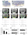
Defects of respiratory rhythm generation in medulla Pre-I neurons of Dscam-deficient mice (C57BL/6 background). A, Parasagittal section of mouse brainstem at E19 stained by Nissl. A dashed line (filled arrow) indicates the anterior limit of the medulla–spinal cord preparation used for the optical imaging (C–K). Another dashed line (open arrow) indicates that used for the measurement of facial (VII) nerve activity (M). B, A magnified image of the squared region in A. A red circle indicates the position of Pre-I neuronal activity, and a blue circle indicates that of Insp neuronal activity, which are shown in C–H. VII, Facial nucleus; LRt, lateral reticular nucleus; nA, nucleus ambiguus; RTN, retrotrapezoid nucleus; SO, superior olive. C–K, The ΔF/F pseudocolor images recorded 500 ms (C–E) or 0 ms (F–H) before C4 inspiratory burst are superimposed on the ventral surface of the medulla. Results of optical imaging are the average of 50 respiratory cycles triggered by C4 inspiratory activity. The red circles indicate the most prominent area of Pre-I neuronal activity, and the blue circles indicate that of Insp neuronal activity. Optical responses within red and blue circles (78 pixels) are quantified and shown as red and blue traces, respectively (I–K). Five mice, analyzed for each genotype, showed the same results and a representative trace from one mouse was shown here. The trace at the bottom (black) shows the C4 ventral root inspiratory activity. Each time point of the images in C–H is shown as vertical dotted lines in the graph (I–K). Note that the Pre-I neuron activity in Dscam−/− mice disappeared (E, K), whereas the activity is moderately retained in Dscam+/− mice (D, J). The preparation was stained with 33 μg/ml Di-2-ANEPEQ for 30 min. The exposure time was 20 ms. Horizontal bar, 0.5 s. The differences of the vertical scales in I–K inevitably derived from the variable conditions, including degrees of staining. L, Areas of Pre-I neuronal activity phase on the traces (I–K) in wild-type (n = 5), Dscam+/− (n = 5), and Dscam−/− (n = 8) mice were calculated by NIH ImageJ 1.33u software. Unit in the ordinate represents the number of pixels that exists in the area. Area of Pre-I neuronal activity in Dscam−/− mice was significantly smaller than that in wild-type mice. *p < 0.05, Tukey–Kramer test compared with wild-type mice. Error bars represent SEM. M, The output of the VII nerve and the C4 ventral root activities in medulla–spinal cord preparations of newborn mice. Horizontal bar under each activity represents a period of 30 s. Vertical bar at the right side of VII nerve or C4 activity represents an amplitude of 0.10 or 0.20 mV, respectively. The VII nerve activities for Dscam−/− became tonic.
Figure 6.
Figure 6.
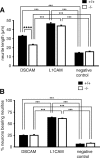
Dscam-dependent neurite outgrowth of DRG neurons. A, B, DRG neurons isolated from wild-type or Dscam−/− E19 mice (C57BL/6 background) were cultured for 7 h on the indicated substrates. The dishes were prepared by sequential coating with poly-d-lysine, anti-Fc antibody, and DSCAM–Fc or L1CAM–Fc chimeric proteins. Dishes coated with poly-d-lysine followed by anti-Fc antibody only were used as negative controls. A, The neurite length of wild-type or Dscam−/− DRG neurons cultured on DSCAM (n = 465 or n = 348), L1CAM (n = 327 or n = 335), or without Fc-fusion protein (negative control) (n = 151 or n = 142) was measured. The neurite length of DRG neurons on DSCAM-coated dish was significantly longer compared with that on negative control dish. ***p < 0.001, Tukey–Kramer test compared with DRG neurons on anti-Fc antibody-coated dish. The neurite length of Dscam−/− DRG neurons on a DSCAM-coated dish was significantly shorter than that of wild-type DRG neurons. ****p < 0.0001, Student's t test compared with wild-type neurons under the same culture conditions. B, The percentage of neurons bearing neurites was quantified. Each bar represents six independent experiments. One hundred neurons were measured for each experiment. The percentage of DRG neurons bearing neurites did not differ significantly between Dscam−/− and wild-type DRG neurons. Statistics on culture conditions with three entities (DSCAM-, L1CAM-, and without Fc-fusion protein-coated dishes) were calculated by Tukey–Kramer test. ***p < 0.001. Error bars represent SEM.
Figure 7.
Figure 7.
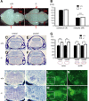
Enlargement of medulla in Dscam-deficient mouse neonates. A, Ventral views of wild-type and Dscam−/− mouse brains at E19 (C57BL/6 background). Arrows indicate the sites for measurement of the width of cerebrum (#) or medulla (##). Dotted lines were provided for comparison. Note that the medulla of Dscam−/− is larger than that of wild-type littermate. B, The averaged width of cerebrum (#) and medulla (##) are 6.05 ± 0.07 and 3.11 ± 0.04 mm, respectively, in wild-type (n = 7) and 6.20 ± 0.07 and 3.35 ± 0.03 mm, respectively, in Dscam−/− mice (n = 7). p = 0.188 for cerebrum and p = 0.0004 (***) for medulla, Student's t test compared with wild type. Error bars represent SEM. Note that the width of medulla of Dscam−/− mice is 7.8% larger than that of wild-type littermates. C–F, Coronal sections containing the rostral (C, D) or the caudal (E, F) part of medulla, including the inferior olive (IO) were stained by Nissl. Although the section for each genotype was prepared at identical anteroposterior positions (rostral end of an inferior olive and ≈150 μm caudal to the rostral section), the sizes for the dorsal structures (cerebellum and inferior colliculus) look different because these were primarily affected by little changes of sectioning angles. The area of medulla (the area marked by a blue or a red dotted line) and the width of medulla (the length marked by a blue or a red arrow) in each of six wild-type or Dscam−/− mice were measured and the values were standardized by wild-type as 100% (G). The medulla of Dscam−/− mice (D, F) is larger in area than that of wild-type mice (C, E). The areas of medulla in Dscam−/− mice were significantly larger than that of wild-type littermates at the rostral (28.7%; ***p = 0.0004) and at the caudal (30.0%; **p = 0.0015) positions. The widths for those were also increased at the rostral (12.0%; **p = 0.0021) and at the caudal (11.2%; *p = 0.0132) positions. Student's t test compared with wild-type, respectively (G). The sizes of cells and their densities in medulla are similar in Dscam−/− and wild type, suggesting that the total cell number is increased in Dscam−/−. H–O, Serial coronal sections from wild-type (H, L) and Dscam−/− (I, M) mice at E19 were stained with Nissl (H–K) and SMI311 antibody recognizing nonphosphorylated neurofilaments, a marker for neuronal soma and dendrites (L–O). Magnified views of boxed areas in H, I, L, or M were represented in J, K, N, or O, respectively. Increased number of Nissl-stained cells in the area dorsolateral to the VII nucleus was indicated by an arrow (K). The numbers of SMI311-immunoreactive cells and fibers are increased in Dscam−/− mice (M, O).
Figure 8.
Figure 8.
Abnormal respiration in Dscam-deficient mice on C57BL/6–BALB/c mixed background. A, Spontaneous respiration activity of newborn (P0) mice was measured by plethysmography. Recordings correspond to spontaneous respiration in wild-type, Dscam+/−, and Dscam−/− littermates, respectively. Horizontal bar under each respiration activity represents a period of 10 s (left side) or 1 s (right side), respectively. Vertical bar at the right side of respiration activity represents a volume of 0.10 mmH2O. Dscam−/− mice show irregular respiratory patterns but milder than those on C57BL/6 mice (Fig. 2). B, C4 ventral root inspiratory activities in medulla–spinal cord preparations of newborn mice on the mixed background. Horizontal bar under each C4 activity represents a period of 1 min. Vertical bar at the right side of C4 activity represents an amplitude of 0.20 mV. The C4 activities for Dscam−/− mice were slow. Notably, low-amplitude C4 activity representing apnea was rarely observed in Dscam−/− mice. For quantifications, see Figure 3.
Figure 9.
Figure 9.
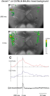
Retained Pre-I neuronal activity in Dscam-deficient mice on C57BL/6–BALB/c mixed background. The ΔF/F pseudocolor images on −350 ms (A) or 0 ms (B) from C4 inspiratory burst are superimposed on the ventral surface of the medulla. Results are the average of 50 respiratory cycles triggered by C4 inspiratory activity. The red circles indicate the most prominent area of Pre-I neuronal activity, and the blue circles indicate that of Insp neuronal activity. Optical responses within red and blue circles (78 pixels) are quantified and shown as red and blue traces, respectively (C). Two Dscam−/− mice showed the same results, and a representative trace from one mouse was shown here. The trace at the bottom (black) shows the C4 ventral root inspiratory activity. Note that the Pre-I neuron activity in Dscam−/− mice on C57BL/6 and BALB/c mixed background is essentially retained. The preparation was stained with 33 μg/ml Di-2-ANEPEQ for 30 min. The exposure time was 20 ms. Horizontal bar, 0.5 s.

References

    1. Agarwala KL, Nakamura S, Tsutsumi Y, Yamakawa K. Down syndrome cell adhesion molecule DSCAM mediates homophilic intercellular adhesion. Brain Res Mol Brain Res. 2000;79:118–126. - PubMed
    1. Agarwala KL, Ganesh S, Amano K, Suzuki T, Yamakawa K. DSCAM, a highly conserved gene in mammals, expressed in differentiating mouse brain. Biochem Biophys Res Commun. 2001;281:697–705. - PubMed
    1. Amiel J, Laudier B, Attié-Bitach T, Trang H, de Pontual L, Gener B, Trochet D, Etchevers H, Ray P, Simonneau M, Vekemans M, Munnich A, Gaultier C, Lyonnet S. Polyalanine expansion and frameshift mutations of the paired-like homeobox gene PHOX2B in congenital central hypoventilation syndrome. Nat Genet. 2003;33:459–461. - PubMed
    1. Arata A, Onimaru H, Homma I. Respiration-related neurons in the ventral medulla of newborn rats in vitro. Brain Res Bull. 1990;24:599–604. - PubMed
    1. Barlow GM, Micales B, Chen XN, Lyons GE, Korenberg JR. Mammalian DSCAMs: roles in the development of the spinal cord, cortex, and cerebellum? Biochem Biophys Res Commun. 2002;293:881–891. - PubMed

Publication types

MeSH terms

LinkOut - more resources