Review
. 2007 Jan-Mar;1(1):19-27.
Epub 2007 Jan 28.

Cell migration from baby to mother

Affiliations
Review

Cell migration from baby to mother

Gavin S Dawe et al. Cell Adh Migr. 2007 Jan-Mar.

Abstract

Fetal cells migrate into the mother during pregnancy. Fetomaternal transfer probably occurs in all pregnancies and in humans the fetal cells can persist for decades. Microchimeric fetal cells are found in various maternal tissues and organs including blood, bone marrow, skin and liver. In mice, fetal cells have also been found in the brain. The fetal cells also appear to target sites of injury. Fetomaternal microchimerism may have important implications for the immune status of women, influencing autoimmunity and tolerance to transplants. Further understanding of the ability of fetal cells to cross both the placental and blood-brain barriers, to migrate into diverse tissues, and to differentiate into multiple cell types may also advance strategies for intravenous transplantation of stem cells for cytotherapeutic repair. Here we discuss hypotheses for how fetal cells cross the placental and blood-brain barriers and the persistence and distribution of fetal cells in the mother.


Figures

Figure 1
Figure 1
A simplified diagrammatic representation of the structure of the human placenta (adapted from Georgiades et al.36) and hypothesized mechanisms of fetomaternal cell traffic. From the end of the first trimester, maternal blood flows into the fetal placenta via the maternal spiral arteries, through the intervillous space bathing the branches of the villous trees and out through the maternal veins (red arrows on left-hand side). The fetal blood enters via the umbilical cord and circulates to the fetal capillaries in the villous trees. A layer of zygote-derived trophoblasts, in humans a syncytium of syncytiotrophoblasts, on the surface of the villous trees (dark green) forms the barrier between the fetal tissues and the maternal blood. Zygote-derived trophoblasts also progressively invade the placental bed and line the maternal vasculature. By the third trimester the maternal spiral arteries are lined through to the (im), while the maternal veins are lined to the border between the decidua basalis (db) and basal plate (bp). In the mouse, the analogue of the fetal placenta is labyrinthine and the trophoblastic invasion of the maternal blood vessels does not extend beyond the junctional zone analogous to the basal plate. Hypothesized mechanisms of fetomaternal cell traffic include (i) deportation of trophoblasts lining the maternal vessels and intervillous space; (ii) microtraumatic hemorrhage; and (iii) cell adhesion and transmigration across the placental barrier.
Figure 2
Figure 2
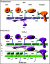
Simplified diagrammatic representations of blood-brain and placental barriers and hypothesized molecular mechanisms of cell adhesion and transmigration. (A) A simplified diagrammatic representation of multistep lymphocyte recognition and capture from blood at the blood brain barrier (adapted from Engelhardt48). Cells expressing α4β1 are captured by VCAM-1 expressed by endothelial cells. There is a rapid activation phase (seconds) that may involve lymphoid chemokines CCL19/ELC and CCL21/SLC. There is a prolonged adhesion phase (hours) followed by slow transmigration (hours) dependent upon binding of LFA-1 to ICAM-1 and/or ICAM-2 on the endothelial cells. It is hypothesized that a similar molecular mechanism may explain fetal cell migration across the blood-brain barrier and the placental barrier. (B) A simplified diagrammatic representation of the human placental barrier showing a hypothetical mechanism of fetal cell capture, adhesion and transmigration. The placental barrier comprises of fetal capillary endothelial cells (fcec), an endothelial basement membrane (ebm), the villous core (vc) which at some interfaces contains pericytes (p) and extracellular matrix, a trophoblastic basement membrane (tbm), in the first trimester a layer of proliferative cytotrophoblasts (ct), and a multinucleated syncytium of syncytiotrophoblasts (ss). In the mouse, the trophoblastic layers differ in that there are two syncytiotrophoblastic layers and the cytotrophoblastic layer is outermost facing the intervillous interface. It is hypothesized that fetal cells may adhere and transmigrate across the placental barrier in a similar manner to that by which lymphocytes cross the blood-brain barrier.
Figure 3
Figure 3
Time course of fetal cell engraftment and persistence in the mouse brain. Adult female mice received intraventricular injection of the excitotoxic NMDA to produce a diffuse brain lesion or were untreated. The mice were crossed with adult male enhanced green fluorescent protein (EGFP) transgenic Green Mice. Fetomaternal microchimerism in the brain was assayed at various time points: gestational days (GD) 7 and 14, the day of parturition (P0), and at seven days (P7), four weeks (P4W) and eight weeks (P8W) post partum (n = 3–8 per group at each time point). The number of fetal cells relative to total cells present in a brain block centered about the site of the injection was quantified by real-time PCR for the EGFP gene in genomic DNA. Procedures were as previously described. There are great individual differences, however, in those mothers in which fetal cells were detected in the brain, the number of fetal cells detected in the brain increases by four weeks post partum and declines again by eight weeks post partum. Overall, in those mothers in which fetal cells persist at four weeks and eight weeks post partum, there are greater numbers of fetal cells in the lesioned brains.

References

    1. Adams KM, Nelson JL. Microchimerism: An investigative frontier in autoimmunity and transplantation. JAMA. 2004;291:1127–1131. - PubMed
    1. Reed W, Lee TH, Norris PJ, Utter GH, Busch MP. Transfusion-associated microchimerism: A new complication of blood transfusions in severely injured patients. Semin Hematol. 2007;44:24–31. - PubMed
    1. Starzl TE. Chimerism and tolerance in transplantation. Proc Natl Acad Sci USA. 2004;101(Suppl 2):14607–14614. - PMC - PubMed
    1. Iverson GM, Bianchi DW, Cann HM, Herzenberg LA. Detection and isolation of fetal cells from maternal blood using the flourescence-activated cell sorter (FACS) Prenat Diagn. 1981;1:61–73. - PubMed
    1. Herzenberg LA, Bianchi DW, Schroder J, Cann HM, Iverson GM. Fetal cells in the blood of pregnant women: Detection and enrichment by fluorescence-activated cell sorting. Proc Natl Acad Sci USA. 1979;76:1453–1455. - PMC - PubMed

LinkOut - more resources