

To adhere or not to adhere: the role of Cadherins in neural crest development
- PMID: 19262148
- PMCID: PMC2637483
- DOI: 10.4161/cam.2.4.6835
To adhere or not to adhere: the role of Cadherins in neural crest development
Abstract
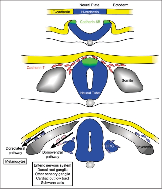
The modulation of cell adhesion is fundamental to the morphogenesis that accompanies proper embryonic development. Cadherins are a large family of calcium-dependent cell adhesion molecules whose spatial and temporal expression is critical to the formation of the neural crest, a unique, multipotent cell type that contributes to the patterning of the vertebrate body plan. Neural crest cells arise from the embryonic ectoderm through inductive interactions and reside in the dorsal aspect of the neural tube. These cells under go an epithelial-to-mesenchymal transition and migrate to precise destinations in the embryo, where they go on to differentiate into such diverse structures as melanocytes, elements of the peripheral nervous system and the craniofacial skeleton. Distinct cadherins are expressed during the induction, migration and differentiation of the neural crest. With the advent of genomic sequencing, assembly and annotation for various model organisms, it has become possible to elucidate the molecular mechanisms underlying cadherin expression, and how these cadherins function, during neural crest development. This review explores the known roles of cadherins and details, where relevant, how different cadherins are regulated during the formation of the neural crest.
Figures

References
-
- Wheelock MJ, Johnson KR. Cadherins as modulators of cellular phenotype. Annu Rev Cell Dev Biol. 2003;19:207–235. - PubMed
-
- Nagar B, Overduin M, Rini JM. Structural basis of calcium-induced E-cadherin rigidification and dimerization. Nature. 1996;380:360–364. - PubMed
-
- Overduin M, Harvery TS, Bagby S, Tong KI, Yau P, Takeichi M, et al. Solution structure of the epithelial cadherin domain responsible for selective cell adhesion. Science. 1995;267:386–389. - PubMed
-
- Shapiro L, Fannon AM, Kwong PD, Thompson A, Lehmann MS, Grübel G, et al. Structural basis of cell-cell adhesion by cadherins. Nature. 1995;374:327–337. - PubMed
-
- Peinado H, Portillo F, Cano A. Transcriptional regulation of cadherins during development and carcinogenesis. Int J Dev Biol. 2004;48:365–375. - PubMed
Publication types
MeSH terms
Substances
Grants and funding
LinkOut - more resources
Full Text Sources