

PtdIns(3,4)P2 instigates focal adhesions to generate podosomes
- PMID: 19262173
- PMCID: PMC2679885
- DOI: 10.4161/cam.3.2.7510
PtdIns(3,4)P2 instigates focal adhesions to generate podosomes
Abstract
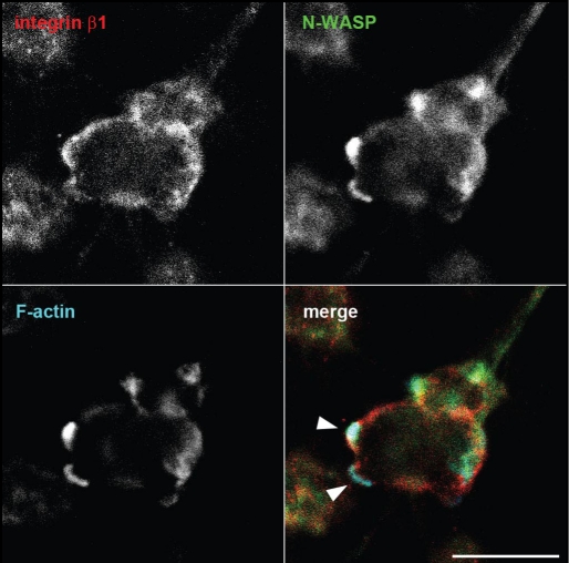
Cell-to-extracellular matrix (ECM) adhesion plays important roles in various biological events, such as proliferation, differentiation and migration. Distinct from other types of adhesion structures (focal complexes, focal adhesions and so on), podosomes and invadopodia are thought to have additional functions beyond attachment, possibly including invasion into the ECM. for podosomes and invadopodia to invade into the ECM, molecules involved in adhesion, actin polymerization and ECM degradation must be recruited to sites of action. Our recent study demonstrated that podosomes form near newly formed focal adhesions via the minimally expressed phosphoinositide PtdIns(3,4) P2-mediated recruitment of the Tks5-Grb2 scaffold, followed by the accumulation of N-WASP. Although this study demonstrated details of molecular interplay during the transformation of focal adhesion, its regulation in the in vivo invasion process remains to be clarified. Here, we discuss the molecular bases of the transformation of focal adhesions to podosomes/invadopodia based on current understanding.
Figures
Comment in
-
Letter From the Guest Editor: New exciting advances in the field of focal adhesion dynamics.Cell Adh Migr. 2009 Apr-Jun;3(2):177-8. doi: 10.4161/cam.3.2.8440. Epub 2009 Apr 16. Cell Adh Migr. 2009. PMID: 19363297 Free PMC article. No abstract available.
References
-
- Gimona M, Buccione R, Courtneidge SA, Linder S. Assembly and biological role of podosomes and invadopodia. Curr Opin Cell Biol. 2008 - PubMed
-
- Marchisio PC, Cirillo D, Teti A, Zambonin-Zallone A, Tarone G. Rous sarcoma virus-transformed fibroblasts and cells of monocytic origin display a peculiar dot-like organization of cytoskeletal proteins involved in microfilament-membrane interactions. Exp Cell Res. 1987;169:202–214. - PubMed
Publication types
MeSH terms
Substances
LinkOut - more resources
Full Text Sources
Research Materials
Miscellaneous