. 2009 May;2(3):220-31.
doi: 10.1038/mi.2009.3. Epub 2009 Mar 4.

MEP1A allele for meprin A metalloprotease is a susceptibility gene for inflammatory bowel disease

Affiliations

MEP1A allele for meprin A metalloprotease is a susceptibility gene for inflammatory bowel disease

S Banerjee et al. Mucosal Immunol. 2009 May.

Abstract

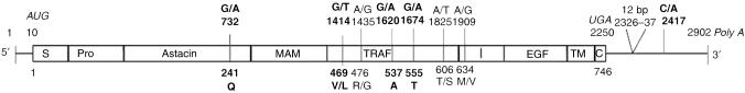
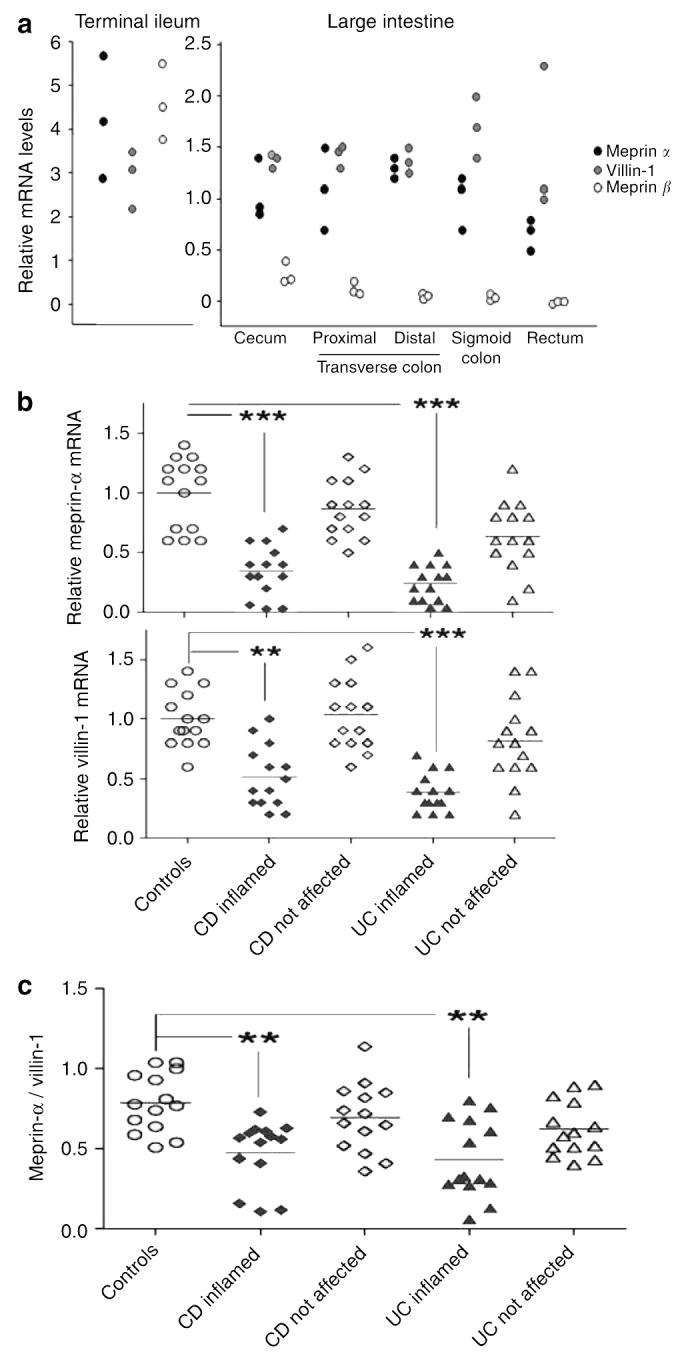
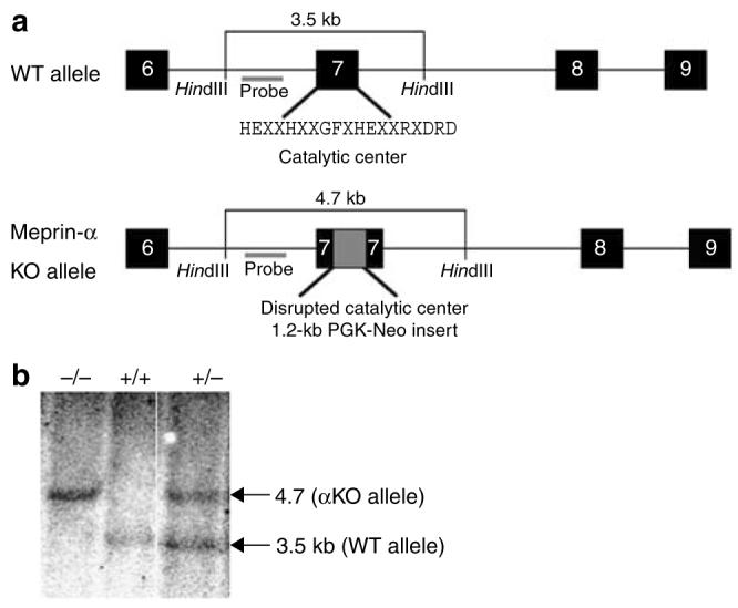
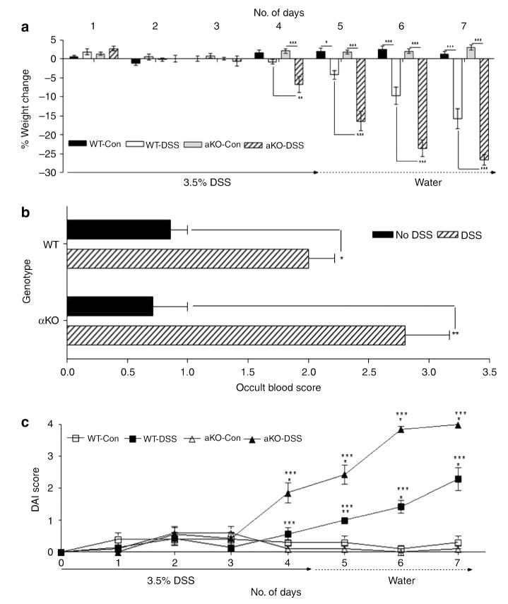
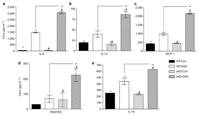
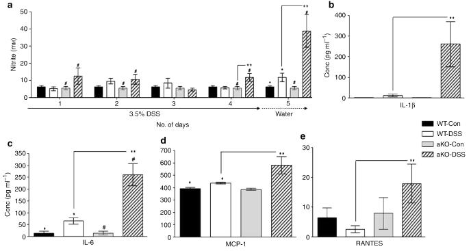
The MEP1A gene, located on human chromosome 6p (mouse chromosome 17) in a susceptibility region for inflammatory bowel disease (IBD), encodes the alpha-subunit of metalloproteinase meprin A, which is expressed in the intestinal epithelium. This study shows a genetic association of MEP1A with IBD in a cohort of ulcerative colitis (UC) patients. There were four single-nucleotide polymorphisms in the coding region (P=0.0012-0.04), and one in the 3'-untranslated region (P=2 x 10(-7)) that displayed associations with UC. Moreover, meprin-alpha mRNA was decreased in inflamed mucosa of IBD patients. Meprin-alpha knockout mice exhibited a more severe intestinal injury and inflammation than their wild-type counterparts following oral administration of dextran sulfate sodium. Collectively, the data implicate MEP1A as a UC susceptibility gene and indicate that decreased meprin-alpha expression is associated with intestinal inflammation in IBD patients and in a mouse experimental model of IBD.


Figures

Figure 1
Figure 1
An overview of MEP1A polymorphisms. The polymorphic sites in the human MEP1A gene are shown in the context of the multidomain structure of the meprin-α subunit. Meprin protein domains are S, signal peptide; Pro, propeptide; Astacin, catalytic protease domain; MAM, meprin/A5-protein/protein-tyrosine phosphate μ; TRAF, TNF-α receptor-associated factor; I, inserted; EGF, epidermal growth factor; TM, transmembrane; and C, cytosolic tail. The AUG start and UGA stop codons and Poly A are italicized. The SNPs and the 12-bp insert are indicated by their nucleotide positions. Affected amino-acid residues and their position in the protein sequence are shown below the subunit structure. UC-associated SNPs are highlighted in bold. Reference mRNA sequence, GenBank NM_005588, which lacks the 12-bp insert. SNP, single-nucleotide polymorphism; TNF, tumor necrosis factor; UC, ulcerative colitis.
Figure 2
Figure 2
Expression of meprin-α, meprin-β, and villin-1 in the intestinal mucosa of IBD patients and healthy controls. mRNA levels, as determined by quantitative RT-PCR, are shown relative to the average colonic expression levels of meprin-α in healthy controls. (a) Endoscopic biopsies of three control individuals collected from the terminal ileum, cecum, proximal and distal transverse colon, sigmoid colon, and rectum. (b) Endoscopic biopsies from the sigmoid colon from not affected and inflamed mucosal areas of CD patients, UC patients, and healthy controls (n=14 in each group). Shown are individual values and mean. ***P<0.001, **P<0.01, compared with controls (Kruskal–Wallis and Dunn's multiple comparison test). (c) Ratio of meprin-α/villin-1 mRNA levels in mucosa in CD and UC patients as compared with controls. IBD, inflammatory bowel disease; CD, Crohn's disease; RT-PCR, reverse transcription PCR; UC, ulcerative colitis.
Figure 3
Figure 3
Strategy for Mep1a gene disruption on mouse chromosome 17. (a) Schematic diagram of a portion of the exon–intron structure of the WT and meprin αKO alleles. Exons (6–9) are represented as black boxes. The neomycin cassette derived from the targeting vector (Osdupdel: gift of O. Smithies) is depicted as a gray box in exon 7 of the KO allele. The 19-amino-acid consensus sequence for the catalytic center of astacin family metalloproteases is also shown. (b) Southern blot analysis of tail-derived genomic DNA. The probe used for Southern blotting detects two HindIII-generated fragments: 3.5 kb corresponding to the WT allele and 4.7 kb from the αKO allele. Lanes: −/−, meprin αKO DNA; +/+, WT DNA; +/−, meprin-α heterozygous DNA. WT, wild type.
Figure 4
Figure 4
Meprin αKO mice are more severely affected than WT mice by DSS treatment. (a) Body weight loss in WT and meprin αKO mice was monitored over a 7-day period (n=7 mice per group). The meprin αKO DSS group lost a greater percent of body weight than the WT DSS group over days 4–7 (*P<0.05; **P<0.01; ***P<0.001). (b) Occult blood scores for both WT and meprin αKO DSS groups on day 7 showed significantly higher bleeding than their corresponding control groups (n=7 mice per group; *P<0.01; **P<0.001). (c) Disease activity index (DAI) for the 3.5% DSS colitis model. WT and meprin αKO controls maintained DAI scores of 0–0.5 over the 7-day period. The DAI for the meprin αKO DSS group was significantly increased starting on day 4 (*P<0.001; compared with controls), whereas the WT DSS DAI increased day 5 onwards (**P<0.01; *P<0.001; compared with controls). There was a significant difference in the DAIs of the two DSS-treated groups from days 4 to 7 (***P<0.001). DSS, dextran sulfate sodium; WT, wild type.
Figure 5
Figure 5
Greater colon injury and inflammation in meprin αKO compared with WT mice. (a) Colon shortening at day 7 for the mice treated with DSS. The average colon length for both WT and meprin αKO mice administered 3.5% DSS was shorter than that for their respective controls at day 7 (*P<0.02; #P<0.0001). The meprin αKO DSS group had shorter colons than WT DSS group (n=7 mice per group; **P<0.04). (b) Proximal colons from WT and meprin αKO mice were scored for injury. Representative histological sections from (i) WT control, (ii) meprin αKO control, (iii) WT DSS-treated, and (iv) meprin αKO DSS-treated colons are shown. Colon sections from both the control groups have normal appearance (i and ii). DSS-treated colon of WT mouse (iii) had crypt destruction (red arrow) along with leukocytic infiltration in the lamina propria as well as in the submucosa (black arrows). Greater damage is evident in the DSS-treated αKO section (iv) in which massive crypt destruction is seen in an area of ulceration (red arrow). Heavy leukocyte infiltration is evident in the lamina propria and submucosal regions (black arrows). (c) The injury scores of WT and meprin αKO DSS-treated groups were normalized to the corresponding control populations. Colons from meprin αKO mice show significantly greater damage than those from WT mice (n=7 mice per group; *P<0.04). (d) MPO activities of WT and meprin αKO colons from DSS-treated and -untreated animals were measured from days 1 to 5. The DSS-treated mice had increased MPO activity relative to their controls (n=6; *P<0.00002; #P<0.00005) and the colon MPO activity of DSS-treated meprin αKO mice was significantly greater than that of the DSS-treated WT mice on days 4 and 5 (**P<0.007). DSS, dextran sulfate sodium; MPO, myeloperoxidase; WT, wild type.
Figure 6
Figure 6
Colon cytokine and chemokine levels of WT and αKO mice treated with or without DSS were measured on day 5 (n=5 per group). Both the DSS-treated groups had significant elevations in their cytokine levels compared with their corresponding controls (WT DSS treated vs. control, *P<0.03; αKO DSS treated vs. control, #P<0.05). (a) IL-6 levels were greatly elevated upon DSS treatment, in both WT and αKO mice, to significantly higher levels in the latter (**P<0.0001). (bd) There were significant increases in the levels of IL-12 (**P<0.01), MCP-1 (**P<0.00007), and RANTES (**P<0.02), in the DSS-treated αKO colons compared with those of the DSS-treated WT group. (e) IL-10, an anti-inflammatory cytokine, was also elevated in both genotypes after DSS treatment; meprin αKO colons had significantly higher levels of IL-10 (**P<0.03). DSS, dextran sulfate sodium; MCP-1, monocyte chemotactic protein-1; WT, wild type.
Figure 7
Figure 7
Meprin αKO mice have greater systemic inflammation than WT mice. (a) Serum nitric oxide levels of all the four groups were measured throughout the course of the study from days 1 to 5. Compared with its control group, DSS-treated meprin αKO mice showed significantly higher levels as early as day 1(#P<0.05), whereas in the WT DSS-treated mouse, values were not elevated until day 5 (*P<0.05). The nitrite levels in meprin αKO mice were significantly higher than the WT DSS-treated group on days 4 and 5 (n=6; **P<0.02). (be) The DSS-treated groups had increased levels of serum cytokines compared with their respective water controls (n=7–15 mice per group; WT DSS treated vs. control, *P<0.03; αKO DSS treated vs. control, #P<0.05). IL-1β and IL-6 levels were markedly elevated in αKO compared with WT serum after DSS treatment (**P<0.04 and P<0.001, respectively). DSS-treated αKO mice also had significantly higher levels of MCP-1 (**P<0.05) and RANTES (**P<0.05) than the DSS-treated WT group. DSS, dextran sulfate sodium; IL, interleukin; MCP-1, monocyte chemotactic protein-1; WT, wild type.

References

    1. Xavier RJ, Podolsky DK. Unravelling the pathogenesis of inflammatory bowel disease. Nature. 2007;448:427–434. - PubMed
    1. Ogura Y, et al. A frameshift mutation in NOD2 associated with susceptibility to Crohn's disease. Nature. 2001;411:603–606. - PubMed
    1. Duerr RH, et al. A genome-wide association study identifies IL23R as an inflammatory bowel disease gene. Science. 2006;314:1461–1463. - PMC - PubMed
    1. Hampe J, et al. Linkage of inflammatory bowel disease to human chromosome 6p. Am. J. Hum. Genet. 1999;65:1647–1655. - PMC - PubMed
    1. Jiang W, et al. Fine mapping of MEP1A, the gene encoding the alpha subunit of the metalloendopeptidase meprin, to human chromosome 6P21. Biochem. Biophys. Res. Commun. 1995;216:630–635. - PubMed

Publication types

MeSH terms

LinkOut - more resources