. 2009 Apr 22;28(8):1157-69.
doi: 10.1038/emboj.2009.58. Epub 2009 Mar 5.

Profilin 1 is required for abscission during late cytokinesis of chondrocytes

Affiliations

Profilin 1 is required for abscission during late cytokinesis of chondrocytes

Ralph T Böttcher et al. EMBO J. .

Abstract

Profilins are key factors for dynamic rearrangements of the actin cytoskeleton. However, the functions of profilins in differentiated mammalian cells are uncertain because profilin deficiency is early embryonic lethal for higher eukaryotes. To examine profilin function in chondrocytes, we disrupted the profilin 1 gene in cartilage (Col2pfn1). Homozygous Col2pfn1 mice develop progressive chondrodysplasia caused by disorganization of the growth plate and defective chondrocyte cytokinesis, indicated by the appearance of binucleated cells. Surprisingly, Col2pfn1 chondrocytes assemble and contract actomyosin rings normally during cell division; however, they display defects during late cytokinesis as they frequently fail to complete abscission due to their inability to develop strong traction forces. This reduced force generation results from an impaired formation of lamellipodia, focal adhesions and stress fibres, which in part could be linked to an impaired mDia1-mediated actin filament elongation. Neither an actin nor a poly-proline binding-deficient profilin 1 is able to rescue the defects. Taken together, our results demonstrate that profilin 1 is not required for actomyosin ring formation in dividing chondrocytes but necessary to generate sufficient force for abscission during late cytokinesis.


Figures

Figure 1
Figure 1
Morphology of Col2pfn1 mice. (A) Western blot analysis of profilin 1 and 2 expressions in total protein lysates from newborn control brain, newborn control and Col2pfn1 cartilage. (B) Northern blot analysis of total RNA from control testes, control cartilage and Col2pfn1 cartilage. (C) Whole mount Alcian blue/Alizarin red staining of control and Col2pfn1 mouse skeletons. Newborn Col2pfn1 mice are indistinguishable from controls but are shorter at 4 weeks of age. (D) Length of long bones of control and Col2pfn1 mice at birth and at 4 weeks of age (mean±s.d., n=8/8 newborn, n=5/5 P28; *P⩽0.01, **P⩽0.05 analysed by Mann–Whitney test). (E) Body length of control and Col2pfn1 mice from birth to 16 days of age (n=6/6). (F) Body weight of control and Col2pfn1 mice from birth to 18 days of age (n=5/5).
Figure 2
Figure 2
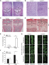
Growth plate cartilage defects in Col2pfn1 mice. (A) Haematoxylin/eosin (H/E)-stained sections of the knee joint region of newborn control and Col2pfn1 mice. (B) H/E-stained sections of the proliferative zone of the tibial epiphyseal growth plate of newborn control and Col2pfn1 mice. (C) H/E-stained sections of the knee joint region of 4-week-old Col2pfn1 and control mice. (D) H/E-stained sections of the tibial epiphyseal growth plate of 4-week-old control and Col2pfn1 mice. Arrows point towards binucleated cells. (E) Quantification of binucleated cells in newborn and 4-week-old growth plates of control and Col2pfn1 mice (mean±s.d., n=3/3, *P<0.05, **P<0.001 analysed by Student's t-test). (F) Quantification of BrdU incorporation (mean±s.d., n=2/3 newborn, n=3/4 P28, *P⩽0.01 analysed by Mann–Whitney test). (G) Sections from the growth plate of 10-day-old control and Col2pfn1 mice stained with fluorescently labelled phalloidin to visualize F-actin. Confocal sections and projections are shown. Abbreviations: r, resting zone; p, proliferative zone; h, hypertrophic zone.
Figure 3
Figure 3
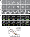
Profilin-deficient cells assemble contractile rings. (AC) Montage of phase-contrast video of mitotic primary control and Col2pfn1 chondrocytes; (A) control chondrocyte, (B) dividing Col2pfn1 chondrocyte and (C) fusing Col2pfn1 chondrocyte. Bar, 10 μm. (D) Montage showing Lifeact localization to the cleavage furrow region of dividing immortalized control and profilin-deficient cells. Selected time points were taken from time-lapse recordings of live cells. Upper panel, confocal section; lower panel, pseudocoloured intensity profile. Time (in minutes and seconds) starts at the onset of anaphase. Bar, 10 μm. (E) Plots of furrow diameter over time after anaphase onset. The furrow diameter is shown for three representative examples of control and profilin-deficient cells.
Figure 4
Figure 4
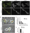
Col2pfn1 chondrocytes fail to complete abscission during late cytokinesis. (A) Control and Col2pfn1 chondrocytes during late cytokinesis stained with an antibody against tubulin and fluorescently labelled phalloidin to visualize F-actin. Bar, 10 μm. (B) Representative deformation maps of control and profilin-deficient cells on a flexible polyacrylamide substrate during cytokinesis. Arrow direction and colour indicate deformation direction and magnitude (blue<green<yellow<red). Bar, 10 μm. (C) Relative distribution of maximal deformation marker displacement around dividing control and profilin-deficient cells (mean±s.d., n=7/7, **P=0.0047 analysed by Mann–Whitney test). (D) Quantification of mean maximal deformation marker displacement around dividing immortalized control and Col2pfn1 cells (mean±s.d., n=7/7, **P=0.0047 analysed by Mann–Whitney test).
Figure 5
Figure 5
Cell spreading and migration are strongly affected in Col2pfn1 chondrocytes. (A) Phase-contrast images of primary newborn control and Col2pfn1 chondrocytes seeded on fibronectin. Selected time points were taken from time-lapse recordings. Col2pfn1 chondrocytes display a spreading defect. (B) Quantification of spreading area of isolated primary control and Col2pfn1 chondrocytes (mean±s.d., summary of four independent control–Col2pfn1 pairs, n>100 cells, ***P⩽0.001 analysed by Mann–Whitney test). (C) Quantification of spreading area of the indicated immortalized cell lines seeded on fibronectin for 24 h (mean±s.d., n>100 cells, NS=not significant, ***P⩽0.001 analysed by Mann–Whitney test). (D) Trajectories of individual primary control and Col2pfn1 cells from the frame-by-frame analysis of time-lapse recordings during a 90-min observation period. The migration velocities of the respective primary cells are indicated (mean±s.d., migration data of over 110 cells from four independent control–Col2pfn1 pairs). (E) Quantification of cell migration velocity of the indicated immortalized cell lines (mean±s.d., migration data of over 140 cells for each cell line from three independent experiments were pooled, NS=not significant, ***P⩽0.001 analysed by Mann–Whitney test). Rescue of profilin-deficient cells with wild-type profilin 1 restores the migratory behaviour.
Figure 6
Figure 6
Profilin function in RhoA-induced actomyosin assembly. (A) Primary control and Col2pfn1 chondrocytes were allowed to spread on fibronectin for 3 h and 12 h and stained with a vinculin antibody (red) and fluorescently labelled phalloidin to visualize F-actin (green). Nuclei were counterstained with DAPI (blue). Confocal sections are shown. Bar, 20 μm. (B) Western blot analysis of total protein lysates from immortalized control and Col2pfn1 cells for the expression of indicated RhoA pathway members. (C) Quantification of RhoA GTP loading in control and profilin-deficient cell lines measured by ELISA (mean±s.d., n=3/3). (D) Western blot analysis of total protein lysates from control and profilin-deficient cell lines for the level of myosin light chain phosphorylation. (E) Western blot analysis of total protein lysates from immortalized Col2pfn1 cells after treatment with the indicated concentrations of ROCK inhibitor Y-27632. (F) Control and profilin-deficient cells were transiently transfected with the indicated constitutively active RhoA pathway members and stained after 16 h with a myc antibody (red) and fluorescently labelled phalloidin to visualize F-actin (green). Nuclei were counterstained with DAPI (blue). Confocal sections are shown. Bar, 20 μm.
Figure 7
Figure 7
Profilin 1 requires actin and poly-proline binding in chondrocytes. (A) Primary Col2pfn1 chondrocytes after viral infection with the indicated profilin 1 variants were allowed to spread on fibronectin for 3 h, fixed and stained with fluorescently labelled phalloidin to visualize F-actin (red). Nuclei were counterstained with DAPI (blue). GFP expression indicates profilin 1-expressing cells. Bar, 20 μm. (B) Western blot analysis of poly-L-proline affinity precipitation from total cell lysates of profilin-deficient cells and cells expressing the indicated profilin 1 variants (profilin 1 wild-type (wt1), profilin 1 H133S (H133S), profilin 1 Y6D (Y6D)).

References

    1. Amano M, Ito M, Kimura K, Fukata Y, Chihara K, Nakano T, Matsuura Y, Kaibuchi K (1996) Phosphorylation and activation of myosin by Rho-associated kinase (Rho-kinase). J Biol Chem 271: 20246–20249 - PubMed
    1. Aszodi A, Chan D, Hunziker E, Bateman JF, Fässler R (1998) Collagen II is essential for the removal of the notochord and the formation of intervertebral discs. J Cell Biol 143: 1399–1412 - PMC - PubMed
    1. Aszodi A, Hunziker EB, Brakebusch C, Fässler R (2003) Beta1 integrins regulate chondrocyte rotation, G1 progression, and cytokinesis. Genes Dev 17: 2465–2479 - PMC - PubMed
    1. Balasubramanian MK, Hirani BR, Burke JD, Gould KL (1994) The Schizosaccharomyces pombe cdc3+ gene encodes a profilin essential for cytokinesis. J Cell Biol 125: 1289–1301 - PMC - PubMed
    1. Betz UA, Vosshenrich CA, Rajewsky K, Müller W (1996) Bypass of lethality with mosaic mice generated by Cre-loxP-mediated recombination. Curr Biol 6: 1307–1316 - PubMed

Publication types

LinkOut - more resources