. 2009;4(3):e4685.
doi: 10.1371/journal.pone.0004685. Epub 2009 Mar 5.

New symmetrically esterified m-bromobenzyl non-aminobisphosphonates inhibited breast cancer growth and metastases

Affiliations

New symmetrically esterified m-bromobenzyl non-aminobisphosphonates inhibited breast cancer growth and metastases

Mohamed Abdelkarim et al. PLoS One. 2009.

Abstract

Background: Although there was growing evidence in the potential use of Bisphosphonates (BPs) in cancer therapy, their strong osseous affinities that contrast their poor soft tissue uptake limited their use. Here, we developed a new strategy to overcome BPs hydrophilicity by masking the phosphonic acid through organic protecting groups and introducing hydrophobic functions in the side chain.

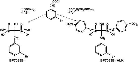
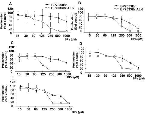
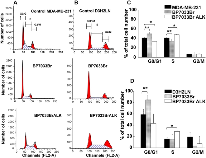
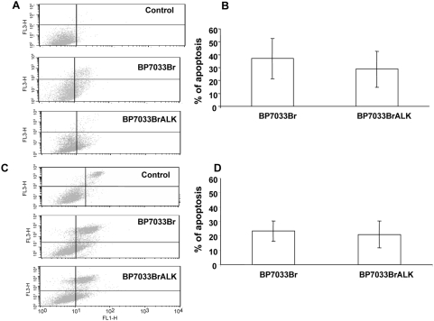
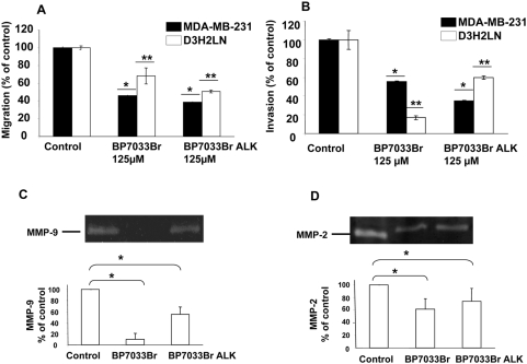
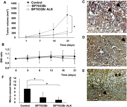
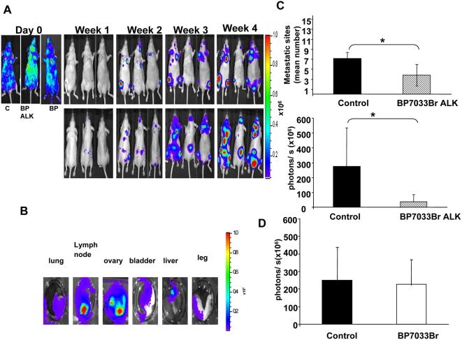
Methodology/principal findings: We synthesized non-nitrogen BPs (non N-BPs) containing bromobenzyl group (BP7033Br) in their side chain that were symmetrically esterified with hydrophobic 4-methoxphenyl (BP7033BrALK) and assessed their effects on breast cancer estrogen-responsive cells (T47D, MCF-7) as well as on non responsive ones (SKBR3, MDA-MB-231 and its highly metastatic derived D3H2LN subclone). BP7033Br ALK was more efficient in inhibiting tumor cell proliferation, migration and survival when compared to BP7033Br. Although both compounds inhibited tumor growth without side effects, only BP7033Br ALK abrogated tumor angiogenesis and D3H2LN cells-induced metastases formation.

Conclusion/significance: Taken together these data suggest the potential therapeutic use of this new class of esterified Bisphosphonates (BPs) in the treatment of tumor progression and metastasis without toxic adverse effects.


Conflict of interest statement

Competing Interests: The authors have declared that no competing interests exist.

Figures

Figure 1
Figure 1. Chemical structure of BP7033Br and BP7033Br ALK.
The first step (1) was an Arbusov reaction between an activated carboxylic function and a very reactive species, the bis(trimethylsilyl) phosphite and the second one (2) was hydrolysis.
Figure 2
Figure 2. BP7033Br and BP7033Br ALK inhibited viability of different breast cancer cells.
T47D (A), MCF-7 (B), SKBR3 (C), MDA-MB-231 (D) or D3H2LN cells (E) (1×10⁵) were treated with BP7033Br and BP7033Br ALK at increasing concentrations for 72 h. Then, the cells were washed and incubated with 0.1 mL of MTT (2 mg/mL) for 4 h. Optical density was measured at 570 nm using a Labsystems Multiskan MS microplate reader. Data represents the mean value (±SD) of three independent experiments.
Figure 3
Figure 3. BP7033Br and BP7033Br ALK inhibited MDA-MB-231 breast cancer cell cycle progression.
Distribution of MDA-MB-231 (A) and D3H2LN (B) cells treated with BPs in different cell cycle phases was determined as described in “Materials and Methods”. Histograms show the percentage of MDA-MB-231 (C) and D3H2LN (D) cell repartition. Each column represents a mean (±SD) of three independent experiments. *P control versus BP7033BrALK and ** P control versus BP7033Br<0.05.
Figure 4
Figure 4. BP7033Br and BP7033Br ALK induced MDA-MB-231 breast cancer cell apoptosis.
Preconfluent MDA-MB-231 (A) and D3H2LN (C) cells were treated with 500 µM BP7033Br or 200 µM BP7033Br ALK for 72 h in a serum-containing medium. Percentages of MDA-MB-231 (B) and D3H2LN (D) were evaluated as described in “Materials and Methods”. Each column represents a mean (±SD) of three independent experiments.
Figure 5
Figure 5. BP7033Br and BP7033Br ALK inhibited MDA-MB-231 breast cancer cell migration, invasion, MMP-9 and MMP-2 activities.
BPs inhibited MDA-MB-231 breast cancer cell migration (A) and invasion (B). Cells (2.5×10⁵) with 125 µM of BPs were added to each 8 µm-insert in the upper chamber of boyden chamber. After 24 h, cells invading the chamber were fixed, stained and counted as described in “Materials and Methods”. BPs inhibited MMP-9 and MMP-2 activities (C and D, respectively). Lyophilized conditioned media were normalized to the number of cells and subjected to 10% SDS-polyacrylamide gels containing 1 mg/mL gelatine. Lane 1, 2 and 3 represent the control, BP7033Br and BP7033Br ALK conditioned medium of treated cells, respectively. Each column represents a mean (±SD) of three independent experiments. *P versus MDA-MB-231 control<0.05, ** P versus D3H2LN control<0.05.
Figure 6
Figure 6. BP7033Br and BP7033Br ALK inhibited D3H2LN tumor growth and esterified analogue completely inhibited angiogenesis.
(A) D3H2LN cells were inoculated in nude mice as described in “Materials and Methods”. After 1 week, mice were treated with BPs (11 mg/kg), twice a week, for 21 days. Each column represents the mean of tumor volume (mm3) (±SD, n = 7). Body weight (BW) ratio was determined for each group (B). Endothelial cells in tumor sections were stained in controls (C), BP7033Br (D) and BP7033Br ALK (E) Microvessels were indicated by arrows and necrosis area by double asterisks (magnification ×100). Quantification of micro-vessel density (F). Each column represents a mean (±SD) of three independent experiments. *P BP7033Br and BP7033Br ALKversus control<0.05.
Figure 7
Figure 7. Only BP7033Br ALK inhibited D3H2LN metastasis.
D3H2LN cells were injected into the left ventricle of nude mice (n = 7). Day 0 showed the successful intracardiac cells injection. Within 2 weeks, when metastasis were initiated, mice were treated with BP7033Br ALK or BP7033Br (A). At the indicated days, the bioluminescence images were acquired for control (c, left panel) and BPs treated mice (BP7033BrALK and BP7033Br middle and right panel, respectively). Ex vivo data confirm soft tissue metastasis from D3H2LN cells injection (B). Quantification of the mean metastatic sites and the photons/s after BP7033Br ALK treatment (C). Quantification the photons/s after BP7033Br treatment (D). Each column represents a mean (±SD) of three independent experiments. *P versus control<0.05.

References

    1. Clezardin P, Ebetino FH, Fournier P. Bisphosphonates and cancer-induced bone disease: beyond their antiresorptive activity. Cancer Res. 2005;65:4971–4974. - PubMed
    1. Clezardin P, Fournier P, Boissier S, Peyruchaud O. In vitro and in vivo antitumor effects of bisphosphonates. Curr Med Chem. 2003;10:173–180. - PubMed
    1. Caraglia M, Santini D, Marra M, Vincenzi B, Tonini G, et al. Emerging anti-cancer molecular mechanisms of aminobisphosphonates. Endocr Relat Cancer. 2006;13:7–26. - PubMed
    1. Green JR. Antitumor effects of bisphosphonates. Cancer. 2003;97:840–847. - PubMed
    1. Rogers MJ. New insights into the molecular mechanisms of action of bisphosphonates. Curr Pharm Des. 2003;9(32):2643–2658. - PubMed

Publication types

MeSH terms