. 2009;4(3):e4671.
doi: 10.1371/journal.pone.0004671. Epub 2009 Mar 5.

Phosphodiesterase 3B is localized in caveolae and smooth ER in mouse hepatocytes and is important in the regulation of glucose and lipid metabolism

Affiliations

Phosphodiesterase 3B is localized in caveolae and smooth ER in mouse hepatocytes and is important in the regulation of glucose and lipid metabolism

Karin Berger et al. PLoS One. 2009.

Abstract

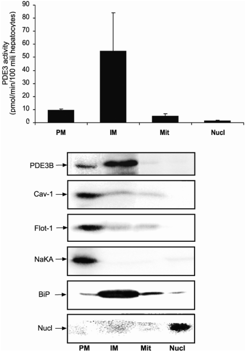
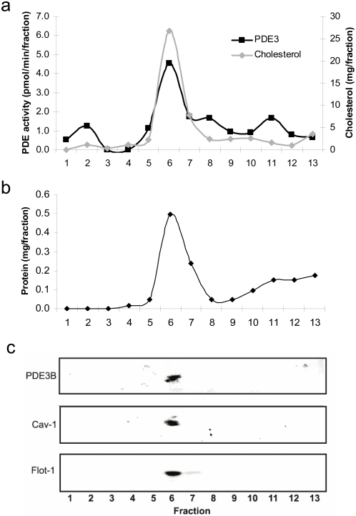
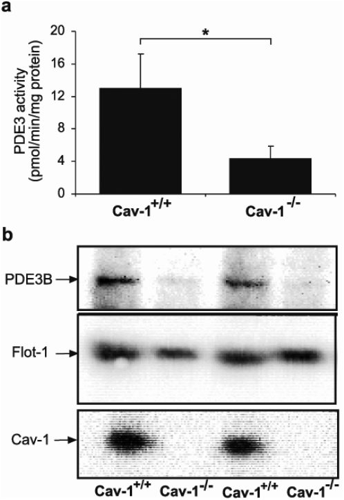
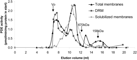
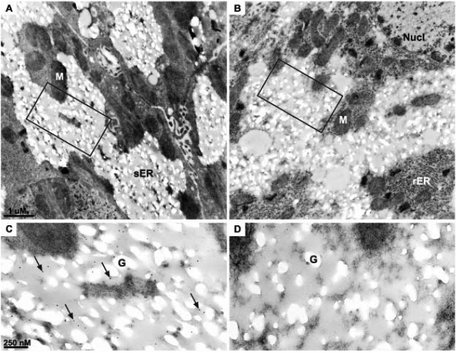
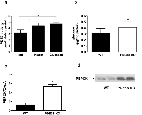
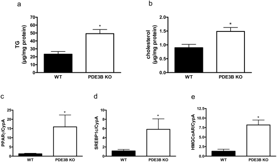
Cyclic nucleotide phosphodiesterases (PDEs) are important regulators of signal transduction processes mediated by cAMP and cGMP. One PDE family member, PDE3B, plays an important role in the regulation of a variety of metabolic processes such as lipolysis and insulin secretion. In this study, the cellular localization and the role of PDE3B in the regulation of triglyceride, cholesterol and glucose metabolism in hepatocytes were investigated. PDE3B was identified in caveolae, specific regions in the plasma membrane, and smooth endoplasmic reticulum. In caveolin-1 knock out mice, which lack caveolae, the amount of PDE3B protein and activity were reduced indicating a role of caveolin-1/caveolae in the stabilization of enzyme protein. Hepatocytes from PDE3B knock out mice displayed increased glucose, triglyceride and cholesterol levels, which was associated with increased expression of gluconeogenic and lipogenic genes/enzymes including, phosphoenolpyruvate carboxykinase, peroxisome proliferator-activated receptor gamma, sterol regulatory element-binding protein 1c and hydroxyl-3-methylglutaryl coenzyme A reductase. In conclusion, hepatocyte PDE3B is localized in caveolae and smooth endoplasmic reticulum and plays important roles in the regulation of glucose, triglyceride and cholesterol metabolism. Dysregulation of PDE3B could have a role in the development of fatty liver, a condition highly relevant in the context of type 2 diabetes.


Conflict of interest statement

Competing Interests: The authors have declared that no competing interests exist.

Figures

Figure 1
Figure 1. Localization of PDE3B in isolated hepatocytes.
Hepatocytes (35–100×106) were isolated from C57BL/6 mice and subjected to subcellular fractionation. Four compartments were isolated; the plasma membranes (PM), internal membranes (IM), mitochondria (Mit) and Nuclei (Nucl). The fractions were subjected to PDE3 activity measurements (a) and immunoblot analysis (b) with antibodies against PDE3B, caveolin-1 (Cav-1, a caveolae marker), flotillin-1 (Flot-1, a lipid raft marker), Na+K+-ATPase (NaKA, a PM marker), BiP (ER marker) and nucleoporin p62 (Nucl, a nucleus marker). PDE3 activities are shown as mean±SEM and immunoblots as one representative experiment (n = 4). Amount of proteins loaded on the gel: PM, 79 µg; IM, 82 µg; Mitochondria, 31 µg; Nuclei, 31 µg.
Figure 2
Figure 2. PDE3B is present in caveolae-enriched fractions of hepatocyte plasma membranes.
Plasma membranes isolated from mouse hepatocytes (50–120×106) were sonicated in NaCO3 and subjected to sucrose gradient fractionation as described in Materials and Methods. Fractions of 0.8 ml were collected starting from the top. The fractions were analyzed for PDE3 activity and cholesterol (a), proteins (b) and subjected to immunoblot analysis (90 µl of each fraction) using antibodies against PDE3B, caveolin-1 (Cav-1) and flotillin-1 (Flot-1) (c). One representative experiment is shown (n = 4).
Figure 3
Figure 3. PDE3B protein is reduced in plasma membranes isolated from livers of caveolin-1 KO mice.
Plasma membranes isolated from WT and caveolin-1 KO livers were analyzed for PDE3 activity (a) and subjected to immunoblot analysis with antibodies against PDE3B, flotillin-1 (flot-1) and caveolin-1 (cav-1) (b). PDE3 activities (mean±SEM) were significantly different in the two groups (p<0.046). One immunoblot (34 µg protein/lane) with two representative experiments are shown (n = 5).
Figure 4
Figure 4. PDE3B is in large size complexes after gelfiltration.
Total membranes were prepared from isolated mouse hepatocytes (20–100×106) and treated with detergent. A portion was used to isolate non-solubilized detergent resistant membranes (DRM) and solubilized membranes. The different membranes were subjected to Superose-6 chromatography. Fractions were collected and analyzed for PDE3 activity. One representative experiment is shown (n = 4).
Figure 5
Figure 5. Transmission electron micrographs of mouse hepatocytes; PDE3B is localized to the smooth ER.
Sections of lowicryl embedded mouse livers with immunogold labeling for PDE3B (A and C). B and D: negative control with omission of the primary antibody. C and D are higher magnifications of the indicated areas in A and B, respectively. Arrows show examples of gold labeling. Representative micrographs are shown (n = 4). G; Glycogen storage M; Mitochondria, Nucl; nucleus, rER; rough endoplasmic reticulum, sER; smooth ER.
Figure 6
Figure 6. Hormonal regulation and glucose production in mouse hepatocytes.
Hepatocytes were isolated from C57BL/6 mice and cultured on Primaria plates. After 18 hours the hepatocytes were incubated for 10 min with 10 nM insulin, 10 nM glucagon or without stimuli (Ctrl). PDE3 activity was measured in total homogenates (a). Values represent mean±SEM (p<0.032 for Insulin, p<0.024 for Glucagon, n = 5). Hepatocytes were isolated from PDE3B KO and WT mice. For glucose production (b) and Western blot analysis of PEPCK (d), the hepatocytes were cultured on Primaria plates over night before the experiment. RNA for PEPCK mRNA expression analysis (c) was isolated from non-cultured hepatocytes. Values are means±SEM. Glucose production: p<0.006, n = 7. PEPCK mRNA: p<0.02, analyzed in duplicate in two independent experiments from two mice of each genotype. PEPCK western blot: one representative experiment is shown, n = 4.
Figure 7
Figure 7. Triglyceride and cholesterol contents are increased in hepatocytes isolated from PDE3B KO mice.
Hepatocytes (a, c–e) and liver (b) from PDE3B KO and WT mice were analyzed with regard to triglyceride (a) and cholesterol (b) content. Furthermore, PPARγ mRNA (c), SREBP1c mRNA (d) and HMG CoA reductase mRNA (e) were analyzed in hepatocytes. Values are means±SEM. Triglyceride content was analyzed as duplicate lipid extractions from four animals of each genotype, p<0.03. Cholesterol content was measured in duplicate in six mice of each genotype, p<0.03. mRNA expressions were analyzed in duplicate in two independent experiments from two mice of each genotype.

References

    1. Conti M, Beavo J. Biochemistry and physiology of cyclic nucleotide phosphodiesterases: essential components in cyclic nucleotide signaling. Annu Rev Biochem. 2007;76:481–511. - PubMed
    1. Degerman E, Manganiello V. Phosphodiesterase 3B; an important regulator of energy homeostasis. In: Beavo, Houslay, Frances, editors. Cyclic Phosphodiesterases in health and disease. Broca Raton, FL USA: CRC Press; 2007. pp. 79–99.
    1. Thompson PE, Manganiello V, Degerman E. Re-discovering PDE3 inhibitors–new opportunities for a long neglected target. Curr Top Med Chem. 2007;7(4):421–36. - PubMed
    1. Choi YH, Ekholm D, Krall J, Ahmad F, Degerman E, et al. Identification of a novel isoform of the cyclic-nucleotide phosphodiesterase PDE3A expressed in vascular smooth-muscle myocytes. Biochem J. 2001;Jan 1;353(Pt 1):41–50. - PMC - PubMed
    1. Nilsson R, Ahmad F, Swärd K, Andersson U, Weston M, et al. Plasma membrane cyclic nucleotide phosphodiesterase 3B (PDE3B) is associated with caveolae in primary adipocytes. Cell Signal. 2006;18(10):1713–21. - PubMed

MeSH terms

LinkOut - more resources