. 2009 Nov;20(11):1823-35.
doi: 10.1007/s00198-009-0882-5. Epub 2009 Mar 5.

Effects of PTH treatment on tibial bone of ovariectomized rats assessed by in vivo micro-CT

Affiliations

Effects of PTH treatment on tibial bone of ovariectomized rats assessed by in vivo micro-CT

J E M Brouwers et al. Osteoporos Int. 2009 Nov.

Abstract

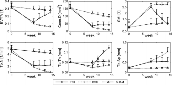
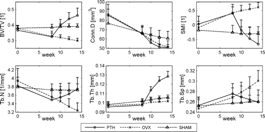
Using in vivo microcomputed tomography (micro-CT), we found in parathyroid hormone (PTH)-treated osteopenic rats linear increases in cortical and trabecular, due to increased trabecular thickness and number, bone mass. Bone was formed in cavities, leading to restoral of nearly cleaved trabeculae. For the first time, effects in PTH-treated rats were analyzed longitudinally.

Introduction: Our aims were to over time (1) determine changes in trabecular thickness and number after PTH, (2) compare responses to PTH between the meta- and epiphysis, (3) determine effects of PTH on mineralization and mechanical properties, (4) determine locations of new bone formation due to PTH on a microlevel, and (5) determine the predictive value of bone structural properties for gain in bone mass after PTH.

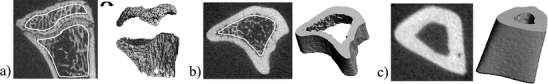
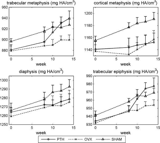
Methods: Adult rats were divided into ovariectomy (OVX; n = 8), SHAM-OVX (n = 8), and OVX and PTH treatment (n = 9). Between weeks 8 and 14, PTH rats received daily subcutaneous PTH injections (60 microg/kg/day). At weeks 0, 8, 10, 12, and 14, in vivo micro-CT scans were made of the proximal and diaphyseal tibia. After sacrifice, all tibiae were tested in three-point bending.

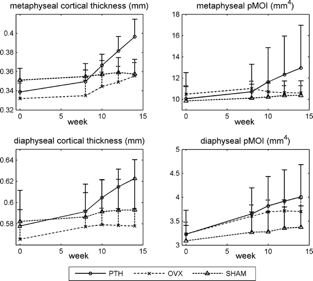
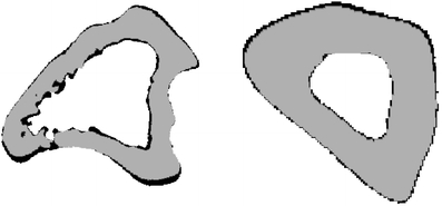
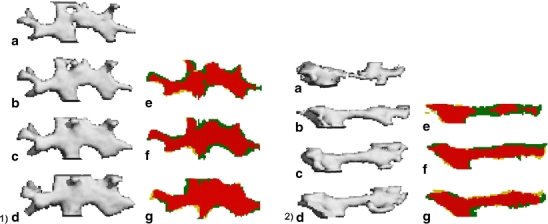
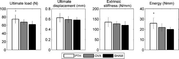
Results: PTH increased bone volume fraction linearly over time in meta- and epiphysis, accompanied by increased trabecular thickness in both and increased trabecular number only in the latter one. CT-estimated mineralization increased in trabecular and remained constant in cortical bone. Ultimate load and energy were increased and ultimate displacement and stiffness unaltered compared to SHAM rats. For those trabeculae analyzed, bone was formed initially on places where it was most beneficial for increasing their strength and later on to all surfaces.


Figures

Fig. 1
Fig. 1
CT scan of a proximal metaphysis showing hand-drawn contours of the metaphyseal and epiphyseal trabecular bone, b proximal metaphysis showing hand-drawn contours of metaphyseal cortical bone, and c diaphyseal cortical bone
Fig. 2
Fig. 2
Structural parameters in the metaphyseal proximal tibia for all groups at all time points (mean ± standard deviation)
Fig. 3
Fig. 3
Structural parameters in the epiphyseal, proximal tibia for all groups at all time points (mean ± standard deviation)
Fig. 4
Fig. 4
Cortical thickness and polar moment of inertia (pMOI) in the meta- and diaphysis of the tibia for all groups at all time points (mean ± standard deviation)
Fig. 5
Fig. 5
Registered images of metaphyseal (left) and diaphyseal (right) cortical bone taken at weeks 8 and 14 showing bone formation during 6 weeks in the cortex of a PTH-treated rat. Gray is bone at week 8, black is newly formed bone
Fig. 6
Fig. 6
CT-estimated bone mineral density of metaphyseal trabecular bone, metaphyseal cortical bone, diaphyseal cortical bone, and epiphyseal trabecular bone (mean ± standard deviation)
Fig. 7
Fig. 7
A trabecula in two PTH-treated ovariectomized rats was tracked over time to determine the development of bone formation (1 and 2). On the left of 1 and 2, you see three-dimensional segmented images of a trabecula, after PTH treatment is started at week 8, taken at weeks 8 (a), 10 (b), 12 (c), and 14 (d). On the right, you see overlaid two-dimensional segmented sections comparing weeks 8 and 10 (e), 10 and 12 (f), and 12 and 14 (g). Yellow indicates resorbed bone, green newly formed bone, and red unchanged bone. Bone formation is clearly seen over time in both trabeculae. In trabecula 1, bone is mostly deposited in the cavities in the first 2 weeks, while later on bone is added to the surface. In trabecula 2, the trabecula appears cleaved after segmentation, although most likely there was still a thin line of bone present. PTH treatment leads to bone formation at the cleaved site, where it is most needed hereby restoring the trabecula
Fig. 8
Fig. 8
Ultimate load, ultimate displacement, extrinsic stiffness, and energy determined from three-point bending test on tibiae after sacrifice at 14 weeks. *p < 0.05 compared to SHAM

References

    1. Mosekilde L, Thomsen JS, McOsker JE. No loss of biomechanical effects after withdrawal of short-term PTH treatment in an aged, osteopenic, ovariectomized rat model. Bone. 1997;20:429–437. doi: 10.1016/S8756-3282(97)00024-0. - DOI - PubMed
    1. Sogaard CH, Mosekilde L, Thomsen JS, Richards A, McOsker JE. A comparison of the effects of two anabolic agents (fluoride and PTH) on ash density and bone strength assessed in an osteopenic rat model. Bone. 1997;20:439–449. doi: 10.1016/S8756-3282(97)00020-3. - DOI - PubMed
    1. Li M, Mosekilde L, Sogaard CH, Thomsen JS, Wronski TJ. Parathyroid hormone monotherapy and cotherapy with antiresorptive agents restore vertebral bone mass and strength in aged ovariectomized rats. Bone. 1995;16:629–635. doi: 10.1016/8756-3282(95)00115-T. - DOI - PubMed
    1. Mosekilde L, Danielsen CC, Sogaard CH, McOsker JE, Wronski TJ. The anabolic effects of parathyroid hormone on cortical bone mass, dimensions and strength—assessed in a sexually mature, ovariectomized rat model. Bone. 1995;16:223–230. doi: 10.1016/8756-3282(94)00033-V. - DOI - PubMed
    1. Mosekilde L, Danielsen CC, Sogaard CH, Thorling E. The effect of long-term exercise on vertebral and femoral bone mass, dimensions, and strength—assessed in a rat model. Bone. 1994;15:293–301. doi: 10.1016/8756-3282(94)90291-7. - DOI - PubMed

Publication types

MeSH terms

Substances