

Manipulation of tissue contrast using contrast agents for enhanced MR microscopy in ex vivo mouse brain
- PMID: 19264139
- PMCID: PMC4441733
- DOI: 10.1016/j.neuroimage.2009.02.027
Manipulation of tissue contrast using contrast agents for enhanced MR microscopy in ex vivo mouse brain
Abstract
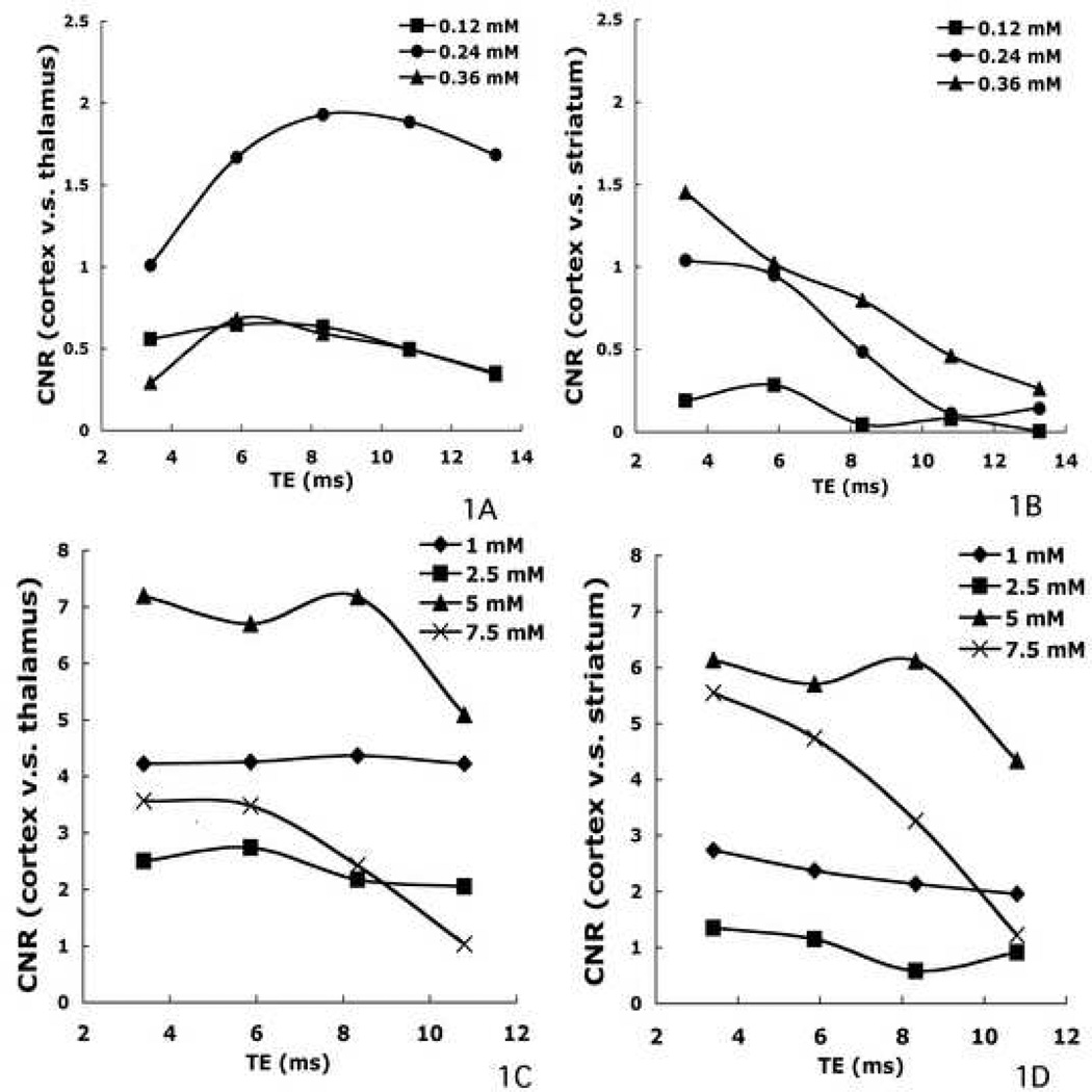
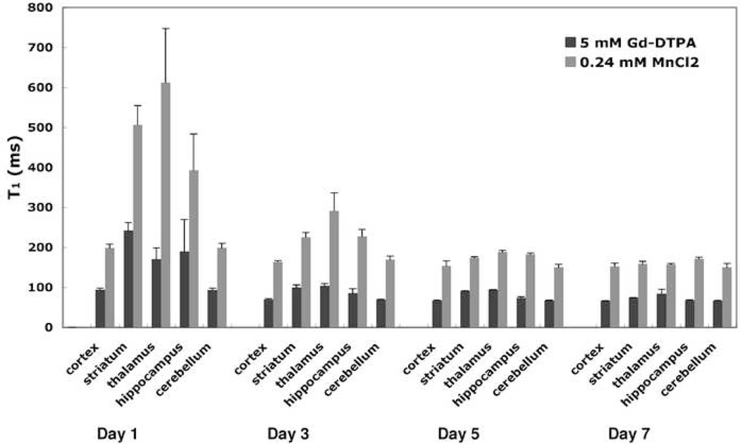
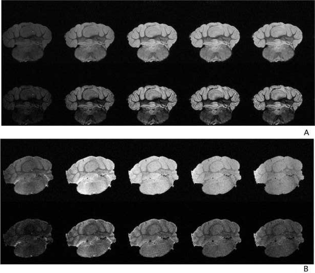
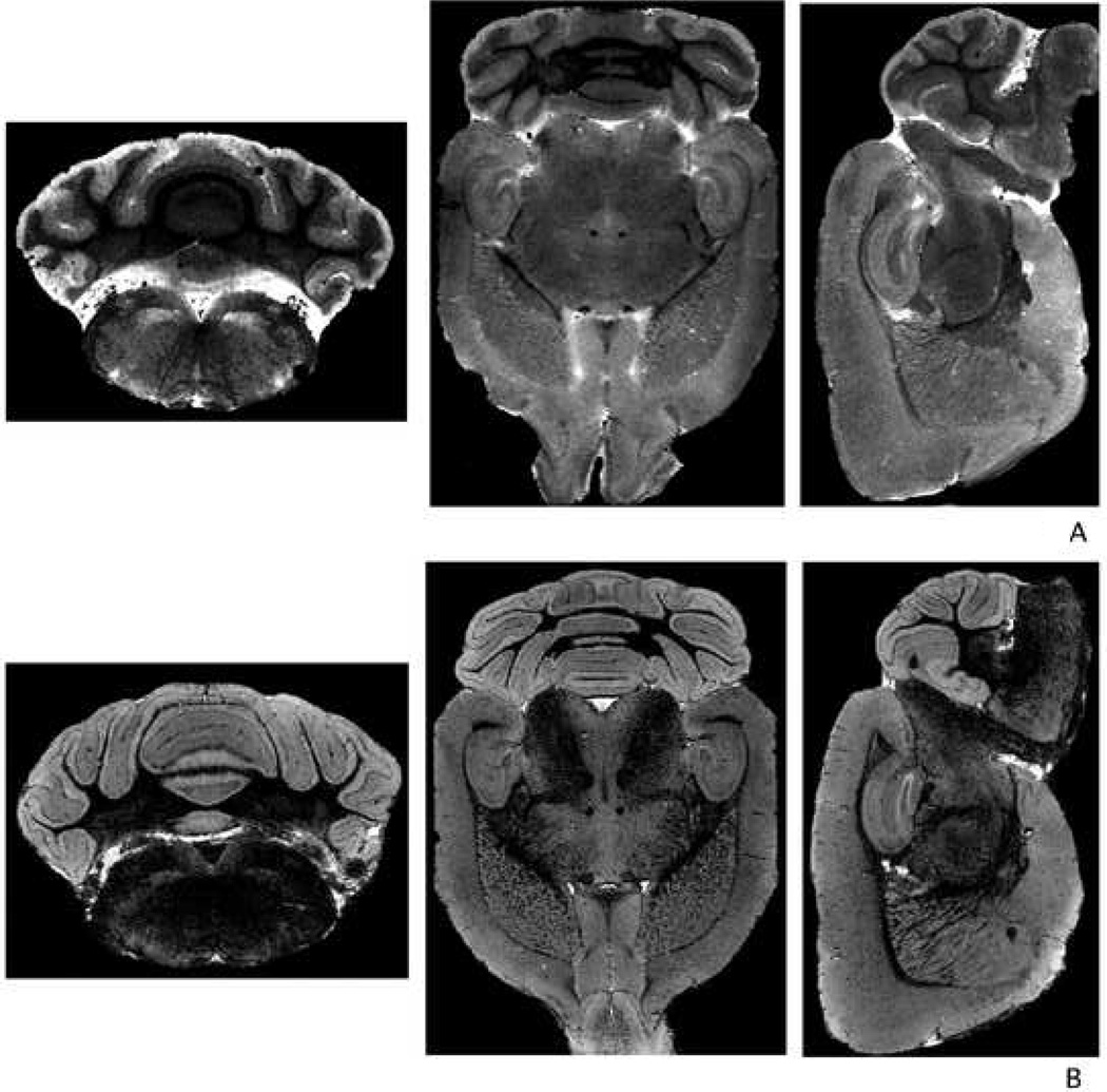
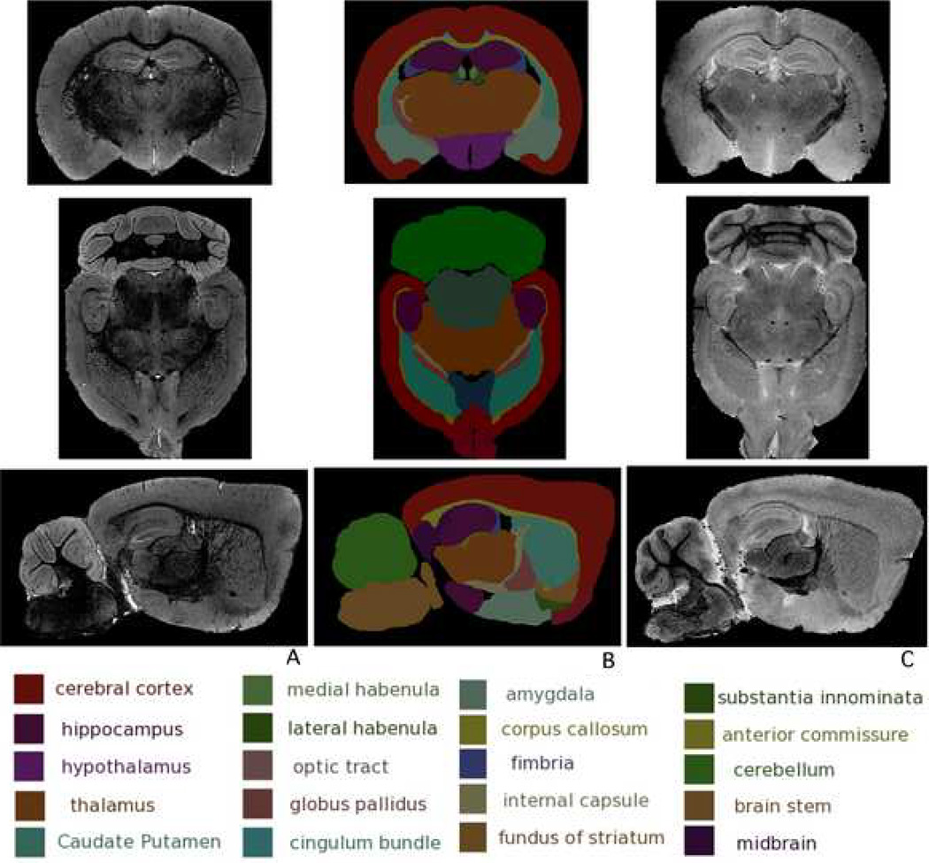
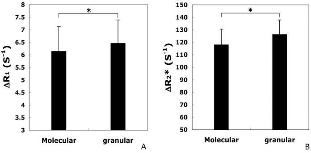
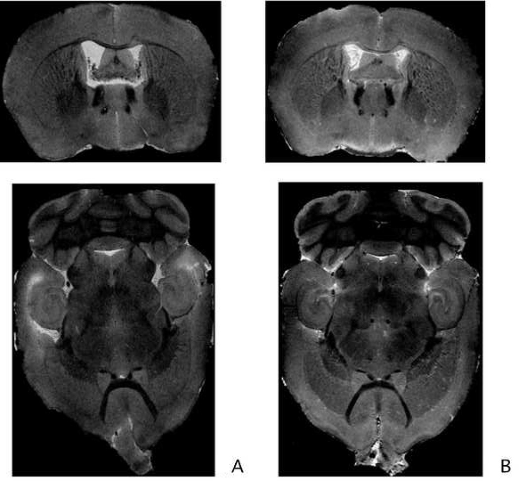
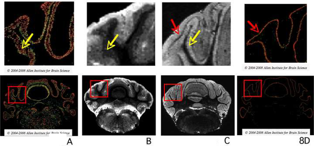
Detailed 3D mouse brain images may promote better understanding of phenotypical differences between normal and transgenic/mutant mouse models. Previously, a number of magnetic resonance microscopy (MRM) studies have successfully established brain atlases, revealing genotypic traits of several commonly used mouse strains. In such studies, MR contrast agents, mainly gadolinium (Gd) based, were often used to reduce acquisition time and improve signal-to-noise ratio (SNR). In this paper, we intended to extend the utility of contrast agents for MRM applications. Using Gd-DTPA and MnCl(2), we exploited the potential use of MR contrast agents to manipulate image contrast by drawing upon the multiple relaxation mechanisms and tissue-dependent staining properties characteristic of each contrast agent. We quantified r(1) and r(2) of Gd-DTPA and MnCl(2) in both aqueous solution and brain tissue and demonstrated the presence of divergent relaxation mechanisms between solution and tissue for each contrast agent. Further analyses using nuclear magnetic resonance dispersion (NMRD) of Mn(2+) in ex vivo tissue strongly suggested macromolecule binding of Mn(2+), leading to increased T(1) relaxation. Moreover, inductively coupled plasma (ICP) mass spectroscopy revealed that MnCl(2) had higher tissue affinity than Gd-DTPA. As a result, multiple regions of the brain stained by the two agents exhibited different image contrasts. Our results show that differential MRM staining can be achieved using multiple MR contrast agents, revealing detailed cytoarchitecture, and may ultimately offer a window for investigating new techniques by which to understand biophysical MR relaxation mechanisms and perhaps to visualize tissue anomalies even at the molecular level.
Figures
Similar articles
-
Structure and Size-selective Permeability of the Synovial Membrane of the Temporomandibular Joint of the Mouse Measured by MR Imaging at 7T.Magn Reson Med Sci. 2015;14(2):115-22. doi: 10.2463/mrms.2014-0058. Epub 2014 Dec 15. Magn Reson Med Sci. 2015. PMID: 25500776
-
Enhanced tissue differentiation in the developing mouse brain using magnetic resonance micro-histology.Magn Reson Med. 2013 Nov;70(5):1380-8. doi: 10.1002/mrm.24573. Epub 2012 Dec 4. Magn Reson Med. 2013. PMID: 23213043
-
Selective visualization of rabbit knee cartilage using MR imaging with a double-contrast agent.J Magn Reson Imaging. 2014 May;39(5):1186-90. doi: 10.1002/jmri.24282. Epub 2013 Sep 30. J Magn Reson Imaging. 2014. PMID: 24123630
-
MR imaging of the breast. Imaging and tissue characterization without intravenous contrast.Magn Reson Imaging Clin N Am. 1994 Nov;2(4):673-90. Magn Reson Imaging Clin N Am. 1994. PMID: 7489316 Review.
-
New Strategies in the Design of Paramagnetic CAs.Contrast Media Mol Imaging. 2020 Sep 27;2020:4327479. doi: 10.1155/2020/4327479. eCollection 2020. Contrast Media Mol Imaging. 2020. PMID: 33071681 Free PMC article. Review.
Cited by
-
Mouse Brain MRI: Including In Vivo, Ex Vivo, and fcMRI for the Study of Microcephaly.Methods Mol Biol. 2023;2583:129-148. doi: 10.1007/978-1-0716-2752-5_12. Methods Mol Biol. 2023. PMID: 36418731
-
Mouse brain fixation to preserve In vivo manganese enhancement for ex vivo manganese-enhanced MRI.J Magn Reson Imaging. 2013 Aug;38(2):482-7. doi: 10.1002/jmri.24005. Epub 2013 Jan 24. J Magn Reson Imaging. 2013. PMID: 23349027 Free PMC article.
-
The brains of aged mice are characterized by altered tissue diffusion properties and cerebral microbleeds.J Transl Med. 2020 Jul 8;18(1):277. doi: 10.1186/s12967-020-02441-6. J Transl Med. 2020. PMID: 32641073 Free PMC article.
-
Deep brain stimulation of the anterior cingulate cortex reduces opioid addiction in preclinical studies.Sci Rep. 2025 Jan 15;15(1):2065. doi: 10.1038/s41598-025-86279-2. Sci Rep. 2025. PMID: 39815019 Free PMC article.
-
Ex vivo mouse brain microscopy at 15T with loop-gap RF coil.Magn Reson Imaging. 2018 Sep;51:1-6. doi: 10.1016/j.mri.2018.04.010. Epub 2018 Apr 18. Magn Reson Imaging. 2018. PMID: 29679634 Free PMC article.
References
-
- Abramoff MD, Magelhaes PJ, Ram SJ. Image Processing with ImageJ. Biophotonics International. 2004;11:36–42.
-
- Aguayo JB, Blackband SJ, Schoeniger J, Mattingly MA, Hintermann M. Nuclear magnetic resonance imaging of a single cell. Nature. 1986;322:190–191. - PubMed
-
- Ali AA, Dale AM, Badea A, Johnson GA. Automated segmentation of neuroanatomical structures in multispectral MR microscopy of the mouse brain. NeuroImage. 2005;27:425–435. - PubMed
-
- Aoki I, Wu Y-JL, Silva AC, Lynch RM, Koretsky AP. In vivo detection of neuroarchitecture in the rodent brain using manganese-enhanced MRI. NeuroImage. 2004;22:1046–1059. - PubMed
-
- Aschner M, Aschner JL. Manganese neurotoxicity: Cellular effects and blood-brain barrier transport. Neuroscience & Biobehavioral Reviews. 1991;15:333–340. - PubMed
Publication types
MeSH terms
Substances
Grants and funding
LinkOut - more resources
Full Text Sources
Medical
Research Materials
