

Prior MDMA (Ecstasy) use is associated with increased basal ganglia-thalamocortical circuit activation during motor task performance in humans: an fMRI study
- PMID: 19264142
- PMCID: PMC2805435
- DOI: 10.1016/j.neuroimage.2009.02.029
Prior MDMA (Ecstasy) use is associated with increased basal ganglia-thalamocortical circuit activation during motor task performance in humans: an fMRI study
Abstract
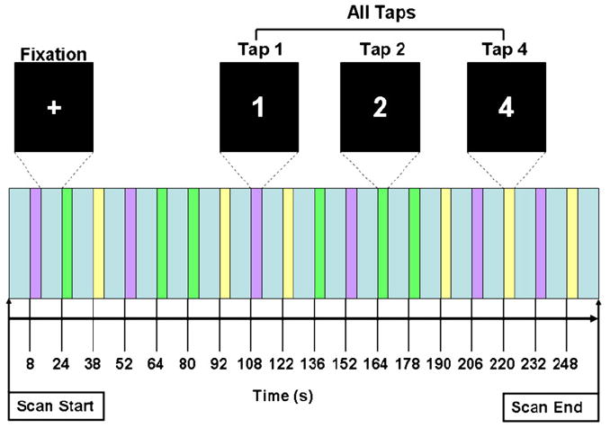
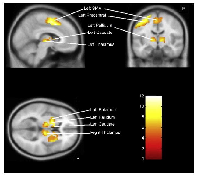
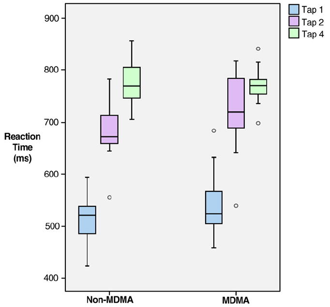
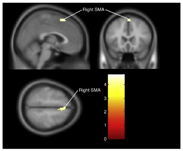
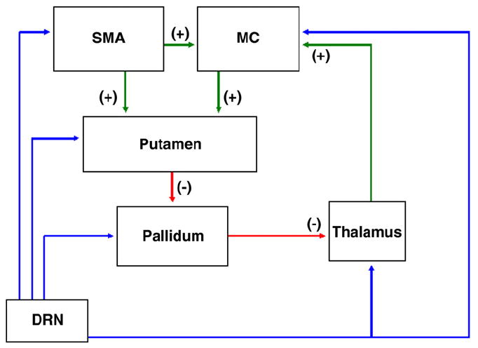
MDMA (3,4-methylenedioxymethamphetamine; Ecstasy) is a popular recreational drug that produces long-lasting serotonin (5-HT) neurotoxicity consisting of reductions in markers for 5-HT axons. 5-HT innervates cortical and subcortical brain regions mediating motor function, predicting that MDMA users will have altered motor system neurophysiology. We used functional magnetic resonance imaging (fMRI) to assay motor task performance-associated brain activation changes in MDMA and non-MDMA users. 24 subjects (14 MDMA users and 10 controls) performed an event-related motor tapping task (1, 2 or 4 taps) during fMRI at 3 T. Motor regions of interest were used to measure percent signal change (PSC) and percent activated voxels (PAV) in bilateral motor cortex, sensory cortex, supplementary motor area (SMA), caudate, putamen, pallidum and thalamus. We used SPM5 to measure brain activation via three methods: T-maps, PSC and PAV. There was no statistically significant difference in reaction time between the two groups. For the Tap 4 condition, MDMA users had more activation than controls in the right SMA for T-score (p=0.02), PSC (p=0.04) and PAV (p=0.03). Lifetime episodes of MDMA use were positively correlated with PSC for the Tap 4 condition on the right for putamen and pallidum; with PAV in the right motor and sensory cortex and bilateral thalamus. In conclusion, we found a group difference in the right SMA and positive dose-response association between lifetime exposure to MDMA and signal magnitude and extent in several brain regions. This evidence is consistent with MDMA-induced alterations in basal ganglia-thalamocortical circuit neurophysiology and is potentially secondary to neurotoxic effects on 5-HT signaling. Further studies examining behavioral correlates and the specific neurophysiological basis of the observed findings are warranted.
Figures
References
-
- Alexander GE, Crutcher MD, DeLong MR. Basal ganglia–thalamocortical circuits: parallel substrates for motor, oculomotor, “prefrontal” and “limbic” functions. Prog Brain Res. 1990;85:119–146. - PubMed
-
- Balogh B, Molnar E, Jakus R, Quate L, Olverman HJ, Kelly PA, Kantor S, Bagdy G. Effects of a single dose of 3,4-methylenedioxymethamphetamine on circadian patterns, motor activity and sleep in drug-naive rats and rats previously exposed to MDMA. Psychopharmacology (Berl) 2004;173:296–309. - PubMed
-
- Bankson MG, Cunningham KA. 3,4-Methylenedioxymethamphetamine (MDMA) as a unique model of serotonin receptor function and serotonin–dopamine interactions. J Pharmacol Exp Ther. 2001;297:846–852. - PubMed
-
- Brett M, Anton JL, Valabregue R, Poline JB. Region of interest analysis using an SPM toolbox 2002
Publication types
MeSH terms
Substances
Grants and funding
LinkOut - more resources
Full Text Sources
Medical
Miscellaneous
