Review
. 2009 Sep;1791(9):949-55.
doi: 10.1016/j.bbalip.2009.02.009. Epub 2009 Mar 2.

Phosphatidic acid signaling to mTOR: signals for the survival of human cancer cells

Affiliations
Review

Phosphatidic acid signaling to mTOR: signals for the survival of human cancer cells

David A Foster. Biochim Biophys Acta. 2009 Sep.

Abstract

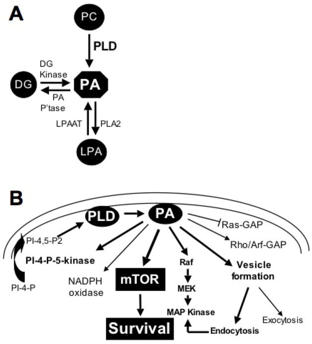
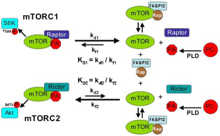
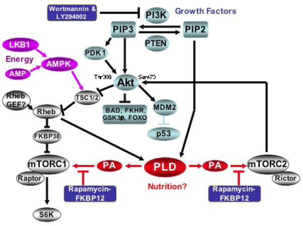
During the past decade elevated phospholipase D (PLD) activity has been reported in virtually all cancers where it has been examined. PLD catalyzes the hydrolysis of phosphatidylcholine to generate the lipid second messenger phosphatidic acid (PA). While many targets of PA signaling have been identified, the most critical target of PA in cancer cells is likely to be mTOR - the mammalian target of rapamycin. mTOR has been widely implicated in signals that suppress apoptotic programs in cancer cells - frequently referred to as survival signals. mTOR exists as two multi-component complexes known as mTORC1 and mTORC2. Recent data has revealed that PA is required for the stability of both mTORC1 and mTORC2 complexes - and therefore also required for the kinase activity of both mTORC1 and mTORC2. PA interacts with mTOR in a manner that is competitive with rapamycin, and as a consequence, elevated PLD activity confers rapamycin resistance - a point that has been largely overlooked in clinical trials involving rapamycin-based strategies. The earliest genetic changes occurring in an emerging tumor are generally ones that suppress default apoptotic programs that likely represent the first line of defense of cancer. Targeting survival signals in human cancers represents a rational anti-cancer therapeutic strategy. Therefore, understanding the signals that regulate PA levels and how PA impacts upon mTOR could be important for developing strategies to de-repress the survival signals that suppress apoptosis. This review summarizes the role of PA in regulating the mTOR-mediated signals that promote cancer cell survival.


Figures

Fig. 1
Fig. 1
Signaling through phosphatidic acid. A. PA is a central node in lipid second messenger signaling. It is generated primarily from phosphatidylcholine (PC) by a hydrolysis reaction that releases the choline head group. PA can also be generated from diacylglycerol (DG) through by a phosphorylation reaction catalyzed by DG kinase; and from lysophosphatidic acid (LPA) via an acylation reaction catalyzed by LPA acyl-transferase (LPAAT). PA can also be converted to both DG and LPA by PA phosphatase (PA P’tase) and a type 2 phospholipase (PLA2). B. PA has many downstream targets. PA has been reported to regulate GTPase activating proteins (GAPs) for Ras, Rho and Arf GTPases; PA also activates an NADPH oxidase [31]. PA activates phosphatidylinositol-4-phosphate (PI-4-P)-5-kinase [31], which may create a positive feedback loop to generate the PI-4,5-bis-phosphate (PI-4,5-P2) required for both PLD1 and PLD2 activity. The likely relevant targets for survival signals provided by PLD and PA are Raf and mTOR, which regulate progression through the cell cycle and suppress apoptosis [32,37]. PA is also required for endocytosis, which along with Raf, contributes to the activation of MAP kinase [73].
Fig. 2
Fig. 2
Model for the differential effects of rapamycin on mTORC1 and mTORC2. The dissociation constants (KD) for mTORC1 and mTORC2 with PA represent the ratios for the rate constants for dissociation (kd) and formation (kf). Recent studies [40] are consistent with a model whereby the rate constant for the dissociation of mTORC1 to PA and mTOR (kd1) is greater than rate constant for the dissociation of mTORC2 to PA and mTOR (kd2). Thus, there are fewer dissociations of PA from mTORC2. The ability of rapamycin-FKBP12 to suppress mTORC1 and mTORC2 is dependent on how frequently mTOR becomes available to bind rapamycin-FKBP12. There would be more mTOR generated from mTORC1 than from mTORC2 and therefore less rapamycin would be required to compete with PA for binding to the mTOR derived from mTORC1. In contrast, the rare dissociations of mTORC2 would require much more rapamycin-FKBP12 to compete with PA to capture the rare mTOR proteins derived from mTORC2. Reducing PA levels would shift the equilibrium in favor of dissociation of the mTOR complexes therefore reduce the concentration of rapamycin-FKBP12 needed to bind to and suppress mTOR.
Fig. 3
Fig. 3
Survival signals generated by PI3K and PLD that target mTOR. In the PI3K pathway, PIP3 recruits Akt and PDK1, a kinase that phosphorylates Akt at Thr308. Akt is also phosphorylated at Ser 473 by mTORC2. Phosphorylation of Akt at Ser473 elevates Akt kinase activity [57] and likely impacts substrate specificity [52]. Akt then phosphorylates several proteins that suppress cell cycle progression – including the TSC1/2 complex, a GTPase activating protein complex that suppresses the GTPase Rheb [53]. Rheb has been reported to stimulate dissociation of FKBP38 from FRB domain of mTOR [55], which contributes to the activation of mTORC1. Rheb was also recently reported to activate PLD1 [38]. PLD-generated PA competes with rapamycin to bind mTORC1 – and possibly FKBP38 – and is required for the mTOR activation of S6 kinase. PLD is also required for the activation of mTORC2, which phosphorylates Akt at Ser473. mTOR is targeted by growth factors, nutrition and energy sensing signals – indicated by different colors.

References

    1. Song J, Jiang YW, Foster DA. EGF induces the production of biologically distinguishable diglyceride species from phosphatidylinositol and phosphatidylcholine: evidence for the independent activation of type C and type D phospholipases. Cell Growth Differ. 1994;5:79–85. - PubMed
    1. Plevin R, Cook SG, Palmer S, Wakelam MJ. Multiple sources of sn-1,2-diacylglycerol in platelet-derived-growth factor-stimulated swiss 3T3 fibroblasts. Biochem J. 1991;279:559–565. - PMC - PubMed
    1. Motoike T, Bieger S, Wiegandt H, Unsicker K. Induction of phosphatidic acid by fibroblast growth factor in cultured baby hamster kidney fibroblasts. FEBS Lett. 1993;332:164–168. - PubMed
    1. Sa G, Das T. Basic fibroblast growth factor stimulates cytosolic phospholipase A2, phospholipase C-γ1 and phospholipase D through distinguishable signaling mechanisms. Mol Cell Biochem. 1999;198:19–30. - PubMed
    1. Karnam P, Standaert ML, Galloway L, Farese RV. Activation and translocation of Rho and ADP ribosylation factor by insulin in rat adipocytes. Apparent involvement of phosphatidylinositol 3-kinase. J Biol Chem. 1997;272:6136–6140. - PubMed

Publication types