Review
. 2009 Feb;20(1):111-8.
doi: 10.1016/j.copbio.2009.02.007. Epub 2009 Mar 4.

Fourier-domain optical coherence tomography: recent advances toward clinical utility

Affiliations
Review

Fourier-domain optical coherence tomography: recent advances toward clinical utility

Brett E Bouma et al. Curr Opin Biotechnol. 2009 Feb.

Abstract

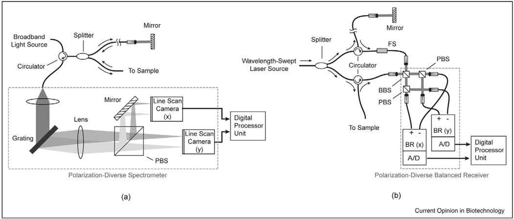
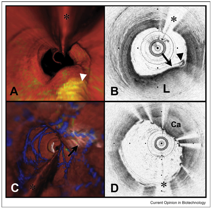
With the advent of Fourier-domain techniques, optical coherence tomography (OCT) has advanced from high-resolution 'point' imaging over small fields-of-view to comprehensive microscopic imaging over three-dimensional volumes that are comparable to the dimensions of luminal internal organs. This advance has required the development of new lasers, improved spectrometers, minimally invasive catheters and endoscopes, and novel optical and signal processing strategies. In recent cardiovascular, ophthalmic, and gastrointestinal clinical studies, the capabilities of Fourier-domain OCT have enabled a new paradigm for diagnostic screening of large tissue areas, which addresses the shortcomings of existing technologies and focal biopsy.


Conflict of interest statement

Conflicts of interest

BEB and GJT: Research sponsored partly by Terumo Corporation, Olympus Corporation, Air Liquide, and Boston Scientific. Co-inventors on patents licensed to LightLab Imaging and Carl Zeiss Meditec, through MIT. Co-inventors on patents licensed to Terumo Corporation and Nidec Corporation, through MGH.

BJV and SHY: Co-inventors on patents licensed to Terumo Corporation and Nidec Corporation, through MGH.

Figures

Figure 1
Figure 1
Schematic layout of typical polarization-diverse Fourier-domain optical coherence tomography systems. (a) Spectral-domain systems rely on a broadband lightsource and a spectrometer as a detector. (b) In frequency-domain systems, the light source is a wavelength-swept laser and the receiver comprises single-element photodiodes. A frequency-shifter is typically used to resolve otherwise degenerate positive and negative depths relative to the reference arm pathlength. PBS: polarization beam splitter; BBS: broadband beam splitter.
Figure 2
Figure 2
Volumetric OFDI imaging of the human LAD coronary artery, obtained in vivo. (A) Fly-through rendering view (proximal-distal) of the OFDI dataset, acquired during a single purge of Lactated Ringers solution (3 ml/s), an imaging catheter pullback rate of 2.0 cm/s and at an image acquisition rate of 100 frames per second. The fly-through depicts a yellow, elevated lipid-rich lesion with scattered macrophages (green). (B) OFDI cross-sectional image obtained at the location of the white arrowhead in (A) demonstrates OFDI evidence of a thin-capped fibroatheroma, a lipid pool (L), a thin cap (black arrow), and a dense band of macrophages at the cap-lipid pool interface. A thin flap of tissue (black arrowhead) can be seen over the cap. (C) Fly-through view (proximal to distal) shows a calcified lesion (black arrow) beneath a newly deployed stent. (D) OFDI cross-sectional image obtained at the location of the black arrow in (C) demonstrates a large calcific nodule (Ca) from 11–5 o’clock causing significant stent distortion, highlighted by the shorter inter-strut distances of the overlying stent compared to the opposing vessel wall. Color scale for (A) and (C): red—artery wall; green—macrophages; yellow—lipid pool; blue—stent. Tick marks, 1 mm. (*) Denotes guide wire artifact.
Figure 3
Figure 3
OFDI images of human Barrett’s esophagus, obtained in vivo. (A) Videoendoscope image shows islands of healthy squamous mucosa intermixed within regions of specialized intestinal metaplasia (SIM). (B) Circular transverse cross-sectional OFDI image with a layered appearance, and in some regions, an irregular surface and intraepithelial glands satisfying the OFDI diagnostic criterion of SIM. The 6 cm longitudinal segment was obtained in approximately 2 min at an acquisition rate of 9.8 frames per second (frame size: 2048 × 4096) and a pullback speed of 0.5 mm/s. (C) Expanded portion of (B) demonstrates surface irregularities (black arrowheads) and glands within the epithelium (red arrowheads). (D) Histopathologic image of the biopsy taken from the involved mucosa shows specialized and non-specialized columnar epithelium, consistent with the OFDI diagnosis of SIM. Scale bars and tick marks represent 1 mm.

References

    1. Huang D, Swanson EA, Lin CP, Schuman JS, Stinson WG, Chang W, Hee MR, Flotte T, Gregory K, Puliafito CA, et al. Optical coherence tomography. Science. 1991;254:1178–1181. - PMC - PubMed
    1. Fercher AF, Hitzenberger CK, Drexler W, Kamp G, Sattmann H. In vivo optical coherence tomography. Am J Ophthalmol. 1993;116:113–115. - PubMed
    1. Tearney GJ, Brezinski ME, Bouma BE, Boppart SA, Pitris C, Southern JF, Fujimoto JG. In vivo endoscopic optical biopsy with optical coherence tomography. Science. 1997;276:2037–2039. - PubMed
    1. Takada K, Yokohama I, Chida K, Noda J. New measurement system for fault location in optical waveguide devices based on interferometric technique. Appl Opt. 1987;26:1603–1606. - PubMed
    1. Youngquist RC, Carr S, Davies DEN. Optical coherence domain reflectometry: a new optical evaluation technique. Opt Lett. 1987;12:158–160. - PubMed

Publication types

MeSH terms