. 2009 May;90(Pt 5):1238-1245.
doi: 10.1099/vir.0.009159-0. Epub 2009 Mar 4.

A truncated T antigen expressed from an alternatively spliced BK virus early mRNA

Affiliations

A truncated T antigen expressed from an alternatively spliced BK virus early mRNA

Johanna R Abend et al. J Gen Virol. 2009 May.

Abstract

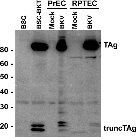
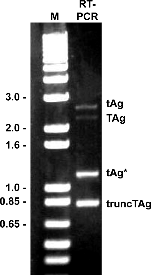
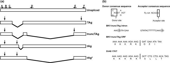
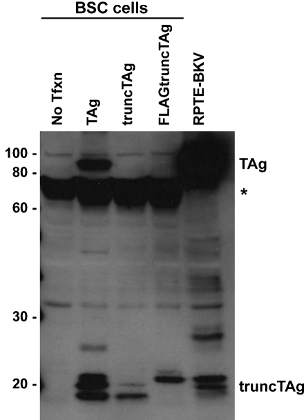
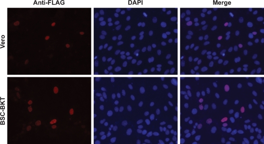
The early region of BK virus (BKV) is known to encode two well-characterized tumour (T) antigens, large T antigen (TAg) and small T antigen (tAg). In this study, we provide evidence of a third early BKV mRNA that codes for an additional early region product with an apparent molecular mass of 17-20 kDa. This truncated form of TAg (truncTAg) is expressed from an alternatively spliced mRNA that is derived from the excision of a second intron from the mRNA encoding TAg. The first 133 aa of truncTAg are identical to those of TAg but the additional splice results in translation from a different reading frame, adding three new amino acids before reaching a stop codon. TruncTAg is expressed in both BKV-transformed and lytically infected cells and it is found to be primarily localized to the nucleus. The function of BKV truncTAg is likely to be relevant to transformation, similar to the additional T antigens of simian virus 40, JC virus and mouse polyomavirus.


Figures

Fig. 1.
Fig. 1.
Detection of truncTAg in whole-cell lysates. Aliquots of total cell protein (30 μg) from BSC and BSC-BKT, mock- or BKV-infected PrECs, or mock- or BKV-infected RPTECs were separated on an 8 % SDS-PAGE gel, transferred to a membrane by Western blot and probed with PAb416. Protein size markers (Magic Mark XP, Invitrogen) are indicated (kDa).
Fig. 2.
Fig. 2.
RT-PCR analysis of BKV early region RNA. RNA was isolated from BSC-BKT cells and used as template in RT-PCR analysis with primers A and F (Supplementary Table S1). The products were analysed on a 0.8 % agarose gel. M, 1 kb Plus Ladder marker (kb; Invitrogen).
Fig. 3.
Fig. 3.
BKV early mRNA structures and truncTAg intron analysis. (a) Diagram of the four early region mRNAs produced by BKV. Boxes indicate exons and lines indicate introns. The unspliced transcript is shown, with the locations of the annealing sites for the primers (Supplementary Table S1) indicated by horizontal arrows. Vertical arrows indicate the positions of the start and stop codons for each transcript. (b) Diagram showing consensus splice donor and acceptor sequences, the DNA sequences of the splice junctions for the truncTAg second intron and the predicted codons adjoining the splice sites. The predicted codons for SV40 17KT are included for comparison.
Fig. 4.
Fig. 4.
Expression of truncTAg from the cDNA clone. BSC cells were transfected with pcDNA3.1(+)-TAg, pcDNA3.1(+)-truncTAg or pcDNA3.1(+)-FLAGtruncTAg, and whole-cell lysates were prepared at 2 days p.t. Aliquots of total cell protein (30 μg) were separated on a 10 % SDS-PAGE gel and analysed by Western blot, probing with PAb416. *, A non-specific band of approximately 70 kDa; No Tfxn, untransfected cells; RPTE-BKV, BKV-infected RPTE whole-cell lysates.
Fig. 5.
Fig. 5.
TruncTAg is localized to the nucleus. Vero (top panels) or BSC-BKT (bottom panels) cells were transfected with pcDNA3.1(+)-FLAGtruncTAg and analysed for localization of FLAG-tagged truncTAg by immunofluorescence, probing with an anti-FLAG epitope antibody as described in Methods. Representative fields of cells are shown at ×200 magnification. Anti-FLAG panels (left) are pseudocoloured in red; areas of colocalization with DAPI appear pink in the merged images (right).

Similar articles

Cited by

References

    1. Abend, J. R., Low, J. A. & Imperiale, M. J. (2007). Inhibitory effect of gamma interferon on BK virus gene expression and replication. J Virol 81, 272–279. - PMC - PubMed
    1. Acott, P. D. & Hirsch, H. H. (2007). BK virus infection, replication and diseases in pediatric kidney transplantation. Pediatr Nephrol 22, 1243–1250. - PMC - PubMed
    1. Ahuja, D., Sáenz-Robles, M. T. & Pipas, J. M. (2005). SV40 large T antigen targets multiple cellular pathways to elicit cellular transformation. Oncogene 24, 7729–7745. - PubMed
    1. Ali, S. H., Kasper, J. S., Arai, T. & DeCaprio, J. A. (2004). Cul7/p185/p193 binding to simian virus 40 large T antigen has a role in cellular transformation. J Virol 78, 2749–2757. - PMC - PubMed
    1. Arthur, A. K., Hoss, A. & Fanning, E. (1988). Expression of simian virus 40 T antigen in Escherichia coli: localization of T-antigen origin DNA-binding domain to within 129 amino acids. J Virol 62, 1999–2006. - PMC - PubMed

Publication types

LinkOut - more resources