. 2009 Jun 1;82(3):439-47.
doi: 10.1093/cvr/cvp083. Epub 2009 Mar 5.

Re-evaluating the efficacy of beta-adrenergic agonists and antagonists in long QT-3 syndrome through computational modelling

Affiliations

Re-evaluating the efficacy of beta-adrenergic agonists and antagonists in long QT-3 syndrome through computational modelling

Rebecca C Ahrens-Nicklas et al. Cardiovasc Res. .

Abstract

Aims: Long QT syndrome (LQTS) is a heterogeneous collection of inherited cardiac ion channelopathies characterized by a prolonged electrocardiogram QT interval and increased risk of sudden cardiac death. Beta-adrenergic blockers are the mainstay of treatment for LQTS. While their efficacy has been demonstrated in LQTS patients harbouring potassium channel mutations, studies of beta-blockers in subtype 3 (LQT3), which is caused by sodium channel mutations, have produced ambiguous results. In this modelling study, we explore the effects of beta-adrenergic drugs on the LQT3 phenotype.

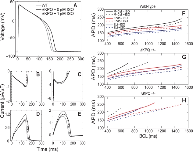
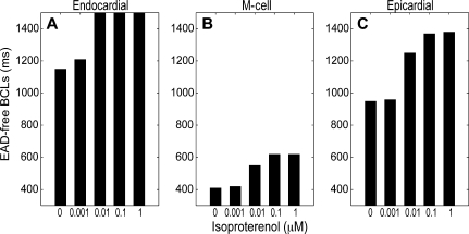
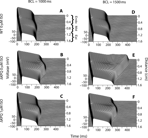
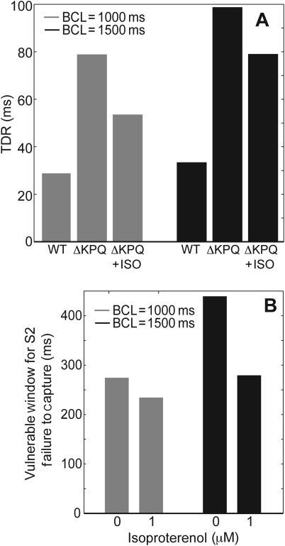
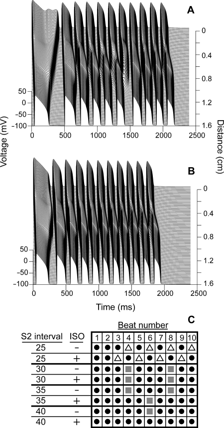
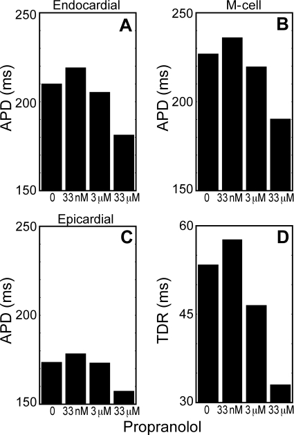
Methods and results: In order to investigate the effects of beta-adrenergic activity and to identify sources of ambiguity in earlier studies, we developed a computational model incorporating the effects of beta-agonists and beta-blockers into an LQT3 mutant guinea pig ventricular myocyte model. Beta-activation suppressed two arrhythmogenic phenomena, transmural dispersion of repolarization and early after depolarizations, in a dose-dependent manner. However, the ability of beta-activation to prevent cardiac conduction block was pacing-rate-dependent. Low-dose beta-blockade by propranolol reversed the beneficial effects of beta-activation, while high dose (which has off-target sodium channel effects) decreased arrhythmia susceptibility.

Conclusion: These results demonstrate that beta-activation may be protective in LQT3 and help to reconcile seemingly conflicting results from different experimental models. They also highlight the need for well-controlled clinical investigations re-evaluating the use of beta-blockers in LQT3 patients.


Figures

Figure 1
Figure 1
Isoproterenol can reverse ΔKPQ mutation-induced action potential durations (APD) prolongation and early after depolarization (EAD) formation. (A–E) Wild-type (grey), ΔKPQ (black), and 1 µM isoproterenol-treated ΔKPQ (dashed) endocardial model myocytes were paced at a basic cycle length (BCL) of 500 ms for 200 beats. APD was prolonged in the mutant cell; this prolongation was partially reversed through treatment with isoproterenol (A). The ΔKPQ mutation causes a late sodium current (B). ICa,L (C), IKr (D), and IKs (E) are all affected by isoproterenol treatment. (F–H) Mid-myocardial (black), endocardial (red), and epicardial (blue) cells were paced with a dynamic restitution protocol in the presence (dashed) and absence (solid) of 1 µM isoproterenol. Wild-type (F), heterozygous ΔKPQ mutant (G), and homozygous ΔKPQ mutant (H) cells were examined. Pacing began at a BCL of 1500 ms and was decreased by 100 ms after 50 beats at each BCL until a final BCL of 400 ms was reached. Only BCLs where no EADs occurred are plotted. Conditions where an EAD occurred were further examined by performing a second pacing protocol that began at the last tested BCL where EADs occurred. A BCL range of 100 ms was then probed in 10 ms increments with 50 beats at each step.
Figure 2
Figure 2
Isoproterenol increases the early after depolarization (EAD)-free BCL window in a dose-dependent manner. Endocardial (A), mid-myocardial (B), and epicardial (C) homozygous ΔKPQ mutant cells were paced with a dynamic restitution protocol as described for Figure 1 in the presence of varying concentrations of isoproterenol. The range of EAD-free BCLs is indicated by the black bars.
Figure 3
Figure 3
Isoproterenol suppresses early after depolarization (EAD) formation and decreases transmural dispersion of repolarization in a mutant model transmural cable. Transmural cardiac cables were paced from the endocardium (top of each panel, position 0 on the cable), and action potentials propagated down through the mid-myocardial and epicardial regions [as labelled in (A)]. A wild-type cable paced at 1000 ms (A) demonstrated shorter action potentials than an untreated ΔKPQ cable (B). One micromolar of isoproterenol reduced ΔKPQ APDs (C). Similar mutation-induced prolongation occurred at a BCL of 1500 ms (E as compared to D), with a non-propagating EAD occurring in the endocardial region. Isoproterenol was able to suppress mutation-induced EADs and APD prolongation (F).
Figure 4
Figure 4
Isoproterenol decreases transmural dispersion of repolarization (TDR) and the vulnerable window for failure to capture by an S2 stimulus. The ΔKPQ mutation increased TDR in a transmural cable. One micromolar of isoproterenol partially reversed this increase (A). The vulnerable window for failure to capture by an S2 stimulus was measured in mutant cables in the presence and absence of isoproterenol at BCLs of 1000 ms (grey) and 1500 ms (black) (B). Failure to capture was defined as a stimulus that failed to evoke an action potential.
Figure 5
Figure 5
The ability of isoproterenol to protect against conduction block depends on the S2 pacing interval. ΔKPQ mutant cables paced at a BCL of 1500 ms were given 10 S2 stimuli at intervals varying from 25 to 40 ms after repolarization of the previous action potential. At an interval of 40 ms, block occurs at the 6th beat in an untreated cable (A) and 1 µM isoproterenol protected against block at this interval (B). The results of experiments at several pacing intervals in the presence and absence of isoproterenol are summarized in (C). Black circles indicate beats where cable-wide propagation occurred; open triangles indicate beats where failure to capture occurred, and grey squares indicate beats where conduction block occurred.
Figure 6
Figure 6
Low-dose propranolol worsens the LQT3 phenotype, but higher doses can decrease both APD and transmural dispersion of repolarization (TDR). Isoproterenol-treated mutant transmural cable models were paced at a BCL of 1000 ms, and treated with 0 µM, 33 nM, 3 µM, or 33 µM propranolol. APD90 in the endocardium (cell 10) (A), mid-myocardium (cell 83) (B), and epicardium (cell 154) (C) were measured. TDR (D) was measured as in Figure 4.

References

    1. Schwartz PJ, Locati EH, Moss AJ, Crampton RS, Trazzi R, Ruberti U. Left cardiac sympathetic denervation in the therapy of congenital long QT syndrome. A worldwide report. Circulation. 1991;84:503–511. - PubMed
    1. Schwartz PJ, Priori SG, Spazzolini C, Moss AJ, Vincent GM, Napolitano C, et al. Genotype-phenotype correlation in the long-QT syndrome: gene-specific triggers for life-threatening arrhythmias. Circulation. 2001;103:89–95. - PubMed
    1. Shimizu W, Noda T, Takaki H, Kurita T, Nagaya N, Satomi K, et al. Epinephrine unmasks latent mutation carriers with LQT1 form of congenital long-QT syndrome. J Am Coll Cardiol. 2003;41:633–642. - PubMed
    1. Moss AJ, Zareba W, Hall WJ, Schwartz PJ, Crampton RS, Benhorin J, et al. Effectiveness and limitations of beta-blocker therapy in congenital long-QT syndrome. Circulation. 2000;101:616–623. - PubMed
    1. Priori SG, Napolitano C, Schwartz PJ, Grillo M, Bloise R, Ronchetti E, et al. Association of long QT syndrome loci and cardiac events among patients treated with beta-blockers. JAMA. 2004;292:1341–1344. - PubMed

Publication types