A yeast glycoprotein shows high-affinity binding to the broadly neutralizing human immunodeficiency virus antibody 2G12 and inhibits gp120 interactions with 2G12 and DC-SIGN
- PMID: 19264785
- PMCID: PMC2682088
- DOI: 10.1128/JVI.02537-08
A yeast glycoprotein shows high-affinity binding to the broadly neutralizing human immunodeficiency virus antibody 2G12 and inhibits gp120 interactions with 2G12 and DC-SIGN
Abstract
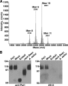
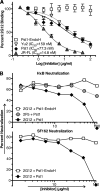
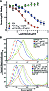
The human immunodeficiency virus type 1 (HIV-1) envelope (Env) protein contains numerous N-linked carbohydrates that shield conserved peptide epitopes and promote trans infection by dendritic cells via binding to cell surface lectins. The potent and broadly neutralizing monoclonal antibody 2G12 binds a cluster of high-mannose-type oligosaccharides on the gp120 subunit of Env, revealing a conserved and highly exposed epitope on the glycan shield. To find an effective antigen for eliciting 2G12-like antibodies, we searched for endogenous yeast proteins that could bind to 2G12 in a panel of Saccharomyces cerevisiae glycosylation knockouts and discovered one protein that bound weakly in a Delta pmr1 strain deficient in hyperglycosylation. 2G12 binding to this protein, identified as Pst1, was enhanced by adding the Delta mnn1 deletion to the Delta pmr1 background, ensuring the exposure of terminal alpha1,2-linked mannose residues on the D1 and D3 arms of high-mannose glycans. However, optimum 2G12 antigenicity was found when Pst1, a heavily N-glycosylated protein, was expressed with homogenous Man(8)GlcNAc(2) structures in Delta och1 Delta mnn1 Delta mnn4 yeast. Surface plasmon resonance analysis of this form of Pst1 showed high affinity for 2G12, which translated into Pst1 efficiently inhibiting gp120 interactions with 2G12 and DC-SIGN and blocking 2G12-mediated neutralization of HIV-1 pseudoviruses. The high affinity of the yeast glycoprotein Pst1 for 2G12 highlights its potential as a novel antigen to induce 2G12-like antibodies.
Figures


Similar articles
-
An engineered Saccharomyces cerevisiae strain binds the broadly neutralizing human immunodeficiency virus type 1 antibody 2G12 and elicits mannose-specific gp120-binding antibodies.J Virol. 2008 Jul;82(13):6447-57. doi: 10.1128/JVI.00412-08. Epub 2008 Apr 23. J Virol. 2008. PMID: 18434410 Free PMC article.
-
Yeast-elicited cross-reactive antibodies to HIV Env glycans efficiently neutralize virions expressing exclusively high-mannose N-linked glycans.J Virol. 2011 Jan;85(1):470-80. doi: 10.1128/JVI.01349-10. Epub 2010 Oct 20. J Virol. 2011. PMID: 20962094 Free PMC article.
-
A glycoconjugate antigen based on the recognition motif of a broadly neutralizing human immunodeficiency virus antibody, 2G12, is immunogenic but elicits antibodies unable to bind to the self glycans of gp120.J Virol. 2008 Jul;82(13):6359-68. doi: 10.1128/JVI.00293-08. Epub 2008 Apr 23. J Virol. 2008. PMID: 18434393 Free PMC article.
-
Antibody responses to the HIV-1 envelope high mannose patch.Adv Immunol. 2019;143:11-73. doi: 10.1016/bs.ai.2019.08.002. Epub 2019 Sep 11. Adv Immunol. 2019. PMID: 31607367 Free PMC article. Review.
-
Sugar and spice: viral envelope-DC-SIGN interactions in HIV pathogenesis.Curr HIV Res. 2003 Jan;1(1):87-99. doi: 10.2174/1570162033352129. Curr HIV Res. 2003. PMID: 15043214 Review.
Cited by
-
A limited number of antibody specificities mediate broad and potent serum neutralization in selected HIV-1 infected individuals.PLoS Pathog. 2010 Aug 5;6(8):e1001028. doi: 10.1371/journal.ppat.1001028. PLoS Pathog. 2010. PMID: 20700449 Free PMC article.
-
Antibodies against Manalpha1,2-Manalpha1,2-Man oligosaccharide structures recognize envelope glycoproteins from HIV-1 and SIV strains.Glycobiology. 2010 Mar;20(3):280-6. doi: 10.1093/glycob/cwp184. Epub 2009 Nov 17. Glycobiology. 2010. PMID: 19920089 Free PMC article.
-
Structural insights into key sites of vulnerability on HIV-1 Env and influenza HA.Immunol Rev. 2012 Nov;250(1):180-98. doi: 10.1111/imr.12005. Immunol Rev. 2012. PMID: 23046130 Free PMC article. Review.
-
Rapid development of glycan-specific, broad, and potent anti-HIV-1 gp120 neutralizing antibodies in an R5 SIV/HIV chimeric virus infected macaque.Proc Natl Acad Sci U S A. 2011 Dec 13;108(50):20125-9. doi: 10.1073/pnas.1117531108. Epub 2011 Nov 28. Proc Natl Acad Sci U S A. 2011. PMID: 22123961 Free PMC article.
-
Broad antiviral activity of carbohydrate-binding agents against the four serotypes of dengue virus in monocyte-derived dendritic cells.PLoS One. 2011;6(6):e21658. doi: 10.1371/journal.pone.0021658. Epub 2011 Jun 30. PLoS One. 2011. PMID: 21738755 Free PMC article.
References
-
- Amberg, D., D. Burke, and J. Strathern. 2005. Methods in yeast genetics: a Cold Spring Harbor Laboratory course manual. Cold Spring Harbor Laboratory Press, Plainview, NY.
-
- Astronomo, R. D., H. K. Lee, C. N. Scanlan, R. Pantophlet, C. Y. Huang, I. A. Wilson, O. Blixt, R. A. Dwek, C. H. Wong, and D. R. Burton. 2008. A glycoconjugate antigen based on the recognition motif of a broadly neutralizing human immunodeficiency virus antibody, 2G12, is immunogenic but elicits antibodies unable to bind to the self glycans of gp120. J. Virol. 826359-6368. - PMC - PubMed
-
- Ballou, C. E. 1990. Isolation, characterization, and properties of Saccharomyces cerevisiae mnn mutants with nonconditional protein glycosylation defects. Methods Enzymol. 185440-470. - PubMed
-
- Bashirova, A. A., T. B. Geijtenbeek, G. C. van Duijnhoven, S. J. van Vliet, J. B. Eilering, M. P. Martin, L. Wu, T. D. Martin, N. Viebig, P. A. Knolle, V. N. KewalRamani, Y. van Kooyk, and M. Carrington. 2001. A dendritic cell-specific intercellular adhesion molecule 3-grabbing nonintegrin (DC-SIGN)-related protein is highly expressed on human liver sinusoidal endothelial cells and promotes HIV-1 infection. J. Exp. Med. 193671-678. - PMC - PubMed
Publication types
MeSH terms
Substances
Grants and funding
LinkOut - more resources
Full Text Sources
Other Literature Sources
Molecular Biology Databases

