. 2009 May;37(8):2539-48.
doi: 10.1093/nar/gkp100. Epub 2009 Mar 5.

Oxidative stress induces degradation of mitochondrial DNA

Affiliations

Oxidative stress induces degradation of mitochondrial DNA

Inna Shokolenko et al. Nucleic Acids Res. 2009 May.

Abstract

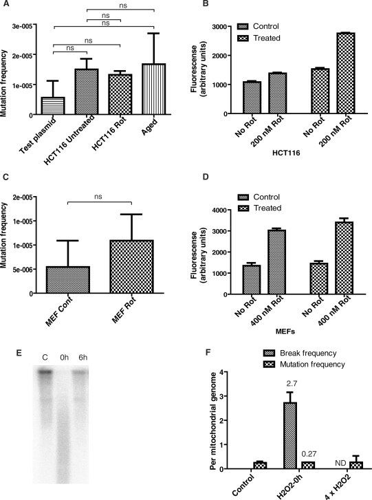
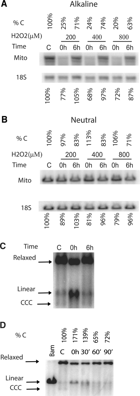
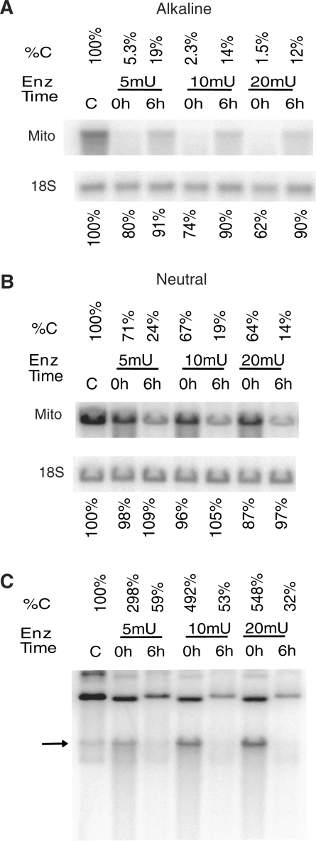
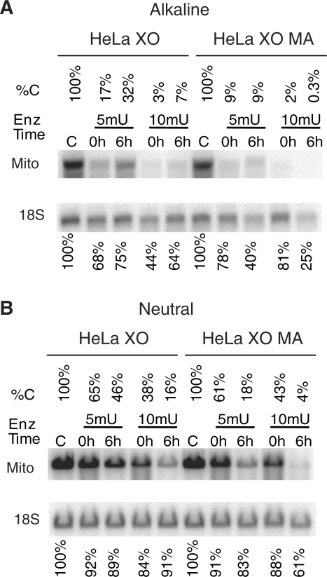
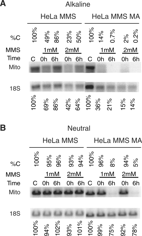
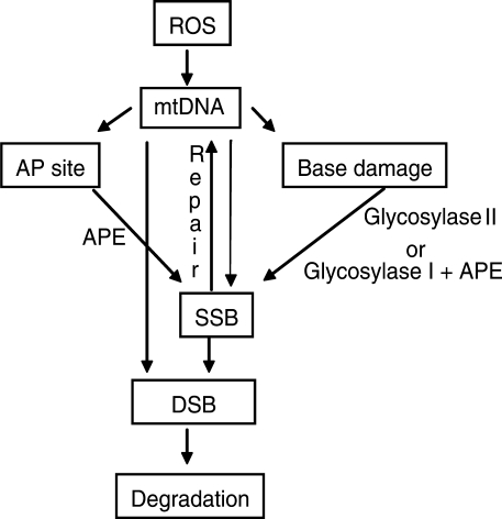
Mitochondrial DNA (mtDNA) is located in close proximity of the respiratory chains, which are the main cellular source of reactive oxygen species (ROS). ROS can induce oxidative base lesions in mtDNA and are believed to be an important cause of the mtDNA mutations, which accumulate with aging and in diseased states. However, recent studies indicate that cumulative levels of base substitutions in mtDNA can be very low even in old individuals. Considering the reduced complement of DNA repair pathways available in mitochondria and higher susceptibility of mtDNA to oxidative damage than nDNA, it is presently unclear how mitochondria manage to maintain the integrity of their genetic information in the face of the permanent exposure to ROS. Here we show that oxidative stress can lead to the degradation of mtDNA and that strand breaks and abasic sites prevail over mutagenic base lesions in ROS-damaged mtDNA. Furthermore, we found that inhibition of base excision repair enhanced mtDNA degradation in response to both oxidative and alkylating damage. These observations suggest a novel mechanism for the protection of mtDNA against oxidative insults whereby a higher incidence of lesions to the sugar-phosphate backbone induces degradation of damaged mtDNA and prevents the accumulation of mutagenic base lesions.


Figures

Figure 1.
Figure 1.
Levels of mtDNA mutations in response to ROS. (A) mtDNA cloned in a plasmid vector (Test plasmid) as well mtDNA from very old individuals (98–101 years old) and mtDNA from HCT116 cells, which were either left untreated or were treated for 30 days with the ROS generator rotenone, were subjected to PCR-cloning-sequencing analysis. No significant increase was detected under any of the conditions tested (n = 3, one-way ANOVA with Tukey post-test). (B and D) Prolonged treatment of HCT116 and MEF cells with rotenone does not result in the selection of cells unable to generate ROS. Control and rotenone-treated (30 days, 200 nM, see ‘Materials and Methods’ section) HCT116 cells were loaded with 5 µM of the mitochondrial superoxide radical indicator MitoSOX, treated with 200 nM rotenone for 30 min, and subjected to FACS analysis; (C) chronic treatment of MEFs with rotenone (400 nM, 30 days) does not cause a significant increase in the mutations frequency (n = 3, two-tailed Student's t-test assuming unequal variances). (E) A representative quantitative Southern blot illustrating both mtDNA damage immediately after treatment (0 h), and repair over 6-h period (6 h). HCT116 cells were treated with 400 µM H2O2 for 60 min, and total BamHI-digested genomic DNA was subjected to both quantitative Southern blotting under the alkaline conditions and PCR-cloning-sequencing. (F) Relative frequency of combined SSBs and abasic sites versus mutagenic base lesions in mtDNA of control (untreated) cells, in mtDNA of cells from (E) (H2O2—0 h), and in mtDNA of cells treated with H2O2 four times with 24 h intervals between treatments (4 × H2O2). The frequency is expressed on a per mtDNA molecule basis (n = 3).
Figure 2.
Figure 2.
Oxidative damage results in the degradation of mtDNA. (A and B) HCT116 cells were either left untreated (C, control) or treated with the indicated concentrations of H2O2 for 1 h, after which total genomic DNA was either extracted immediately (0 h), or allowed to repair for 6 h (6 h) prior to extraction. The DNA was digested with BamHI, which has a single recognition site in human mtDNA, quantitated and separated in 0.6% agarose gel under either alkaline (A) or neutral (B) conditions. After Southern blotting with mitochondrial (Mito) and nuclear (18S) DNA probes, the intensity of bands corresponding to intact mtDNA was expressed as a percent of control (untreated) values. (C) Oxidative DNA damage is accompanied by accumulation of linear mtDNA intermediates. HCT116 cells were treated with 200 µM H2O2, total DNA was extracted, digested with Bgl II, and subjected to Southern blotting under the neutral conditions. Bgl II has no recognition sites in mtDNA, and therefore the banding pattern is reflective of the native state of mtDNA. The positions of markers corresponding to covalently closed circular (CCC), linear and relaxed mtDNA molecules are indicated by arrows. (D) A time course of degradation of linear mtDNA intermediates. HCT116 cells were either left untreated (C) or treated with 400 μM H2O2 for 30 min, and then either lysed immediately, or allowed to recover for 30, 60, or 90 min. The percentage of linear mtDNA as compared to untreated control is indicated.
Figure 3.
Figure 3.
The effect of enzymatically induced oxidative stress on mtDNA. HCT116 cells were either left untreated (C), or treated with 5, 10, or 20 mU of xanthine oxidase (XO) in the presence of hypoxanthine (0.5 mM). Cells were then either lysed immediately after treatment (0 h) or allowed for repair (6 h). Total DNA was extracted and digested with BamHI, and (A), separated under denaturing (alkaline) conditions, and subjected to Southern blotting with nuclear (18S) and mtDNA probe. %C indicates percent intact mtDNA as compared to untreated control. (B) The same samples as in A were run under non-denaturing (TBE buffer) conditions. Annotation is the same as in (A). This blot shows intact and SSBs containing mtDNA and nDNA. (C) Total DNA samples from the same experiment as in (A) and (B) were digested with BglII and run under the non-denaturing conditions to detect linear mtDNA intermediates. Annotation is the same as in (A) and (B). The arrow indicates the position of the linear mtDNA species.
Figure 4.
Figure 4.
Effect of APE1 inhibition on mtDNA degradation in response to oxidative stress. HeLa cells were treated with XO in the presence of hypoxanthine and in the presence or absence of methoxyamine (MA). Cells were then either lysed immediately after treatment (0 h) or allowed for repair (6 h) in the presence or absence of MA. Total DNA was extracted and digested with BamHI, and (A), separated under denaturing (alkaline) conditions, and subjected to Southern blotting with nuclear (18S) and mtDNA (Mito) probes. %C indicates percentage intact mtDNA as compared to untreated control. (B) The same samples as in (A) were run under non-denaturing (TBE buffer) conditions. Annotation is the same as in (A). This blot shows both intact and SSB-containing nDNA and mtDNA.
Figure 5.
Figure 5.
Effect of APE1 inhibition on mtDNA degradation in response to alkylating damage. HeLa cells were treated with indicated concentrations of MMS in the presence or absence of methoxyamine (MA). Cells were then either lysed immediately after treatment (0 h) or allowed for repair (6 h) in the presence or absence of MA. Total DNA was extracted and digested with BamHI, and (A), separated under denaturing (alkaline) conditions, and subjected to Southern blotting with nuclear (18S) and mtDNA (Mito) probes. %C indicates percentage intact mtDNA as compared to untreated control. (B) The same samples as in A were run under non-denaturing (TBE buffer) conditions. Annotation is the same as in (A). This blot shows both intact and SSB-containing nDNA and mtDNA.
Figure 6.
Figure 6.
Proposed interaction between mtDNA repair and degradation pathways. Glycosylase I and Glycosylase II, mono- and bi-functional DNA glycosylases. A bi-functional DNA glycosylase also posseses an AP-lyase activity (makes an incision at an abasic site). APE, apurinic/apyrimidinic endonuclease APE/Ref1. See ‘Discussion’ section for details.

References

    1. Wallace DC, Singh G, Lott MT, Hodge JA, Schurr TG, Lezza AM, Elsas LJ, 2nd., Nikoskelainen EK. Mitochondrial DNA mutation associated with Leber's hereditary optic neuropathy. Science. 1988;242:1427–1430. - PubMed
    1. Chatterjee A, Mambo E, Sidransky D. Mitochondrial DNA mutations in human cancer. Oncogene. 2006;25:4663–4674. - PubMed
    1. Ohta S. Contribution of somatic mutations in the mitochondrial genome to the development of cancer and tolerance against anticancer drugs. Oncogene. 2006;25:4768–4776. - PubMed
    1. Wallace DC, Lott MT, Shoffner JM, Ballinger S. Mitochondrial DNA mutations in epilepsy and neurological disease. Epilepsia. 1994;35(Suppl. 1):S43–S50. - PubMed
    1. Krishnan KJ, Reeve AK, Turnbull DM. Do mitochondrial DNA mutations have a role in neurodegenerative disease? Biochem. Soc. Trans. 2007;35:1232–1235. - PubMed

Publication types