

A nonlinear dynamic model of DNA with a sequence-dependent stacking term
- PMID: 19264801
- PMCID: PMC2673413
- DOI: 10.1093/nar/gkp016
A nonlinear dynamic model of DNA with a sequence-dependent stacking term
Abstract
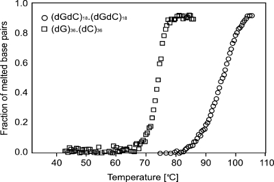
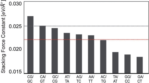
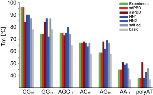
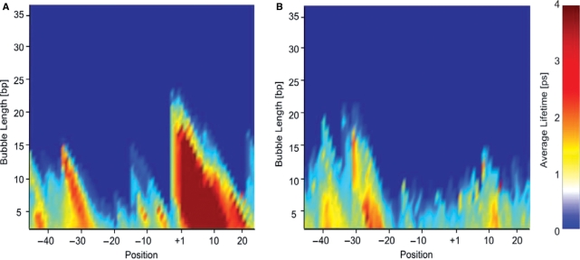
No simple model exists that accurately describes the melting behavior and breathing dynamics of double-stranded DNA as a function of nucleotide sequence. This is especially true for homogenous and periodic DNA sequences, which exhibit large deviations in melting temperature from predictions made by additive thermodynamic contributions. Currently, no method exists for analysis of the DNA breathing dynamics of repeats and of highly G/C- or A/T-rich regions, even though such sequences are widespread in vertebrate genomes. Here, we extend the nonlinear Peyrard-Bishop-Dauxois (PBD) model of DNA to include a sequence-dependent stacking term, resulting in a model that can accurately describe the melting behavior of homogenous and periodic sequences. We collect melting data for several DNA oligos, and apply Monte Carlo simulations to establish force constants for the 10 dinucleotide steps (CG, CA, GC, AT, AG, AA, AC, TA, GG, TC). The experiments and numerical simulations confirm that the GG/CC dinucleotide stacking is remarkably unstable, compared with the stacking in GC/CG and CG/GC dinucleotide steps. The extended PBD model will facilitate thermodynamic and dynamic simulations of important genomic regions such as CpG islands and disease-related repeats.
Figures
Similar articles
-
Multifractal analysis of thermal denaturation based on the Peyrard-Bishop-Dauxois model.Phys Rev E Stat Nonlin Soft Matter Phys. 2011 Sep;84(3 Pt 1):031918. doi: 10.1103/PhysRevE.84.031918. Epub 2011 Sep 19. Phys Rev E Stat Nonlin Soft Matter Phys. 2011. PMID: 22060414
-
Fast Prediction of DNA Melting Bubbles Using DNA Thermodynamic Stability.IEEE/ACM Trans Comput Biol Bioinform. 2015 Sep-Oct;12(5):1137-45. doi: 10.1109/TCBB.2015.2396057. IEEE/ACM Trans Comput Biol Bioinform. 2015. PMID: 26451825
-
Bubble nucleation and cooperativity in DNA melting.Phys Rev Lett. 2005 Jan 28;94(3):035504. doi: 10.1103/PhysRevLett.94.035504. Epub 2005 Jan 27. Phys Rev Lett. 2005. PMID: 15698282
-
Fluctuations in the DNA double helix: A critical review.Phys Life Rev. 2014 Jun;11(2):153-70. doi: 10.1016/j.plrev.2014.01.005. Epub 2014 Jan 13. Phys Life Rev. 2014. PMID: 24560595 Review.
-
DNA melting and energetics of the double helix.Phys Life Rev. 2018 Aug;25:1-21. doi: 10.1016/j.plrev.2017.11.012. Epub 2017 Nov 14. Phys Life Rev. 2018. PMID: 29170011 Review.
Cited by
-
Nonlinear physics opens a new paradigm for accurate transcription start site prediction.BMC Bioinformatics. 2022 Dec 30;23(1):565. doi: 10.1186/s12859-022-05129-4. BMC Bioinformatics. 2022. PMID: 36585618 Free PMC article.
-
DNA dynamics play a role as a basal transcription factor in the positioning and regulation of gene transcription initiation.Nucleic Acids Res. 2010 Apr;38(6):1790-5. doi: 10.1093/nar/gkp1084. Epub 2009 Dec 17. Nucleic Acids Res. 2010. PMID: 20019064 Free PMC article.
-
Bubble Relaxation Dynamics in Homopolymer DNA Sequences.Molecules. 2023 Jan 20;28(3):1041. doi: 10.3390/molecules28031041. Molecules. 2023. PMID: 36770707 Free PMC article.
-
Mammalian stem cells reprogramming in response to terahertz radiation.PLoS One. 2010 Dec 31;5(12):e15806. doi: 10.1371/journal.pone.0015806. PLoS One. 2010. PMID: 21209821 Free PMC article.
-
Evaluating the role of coherent delocalized phonon-like modes in DNA cyclization.Sci Rep. 2017 Aug 29;7(1):9731. doi: 10.1038/s41598-017-09537-y. Sci Rep. 2017. PMID: 28851939 Free PMC article.
References
-
- McClare CW. A quantum mechanical muscle model. Nature. 1972;240:88–90. - PubMed
-
- Davydov AS. The theory of contraction of proteins under their excitation. J. Theor. Biol. 1973;38:559–569. - PubMed
-
- Peyrard M, Bishop AR. Statistical mechanics of a nonlinear model for DNA denaturation. Phys. Rev. Lett. 1989;62:2755–2758. - PubMed
-
- Dauxois T, Peyrard M, Bishop AR. Entropy-driven DNA denaturation. Phys. Rev. E Stat. Phys. Plasmas Fluids Relat. Interdiscip. Topics. 1993;47:R44–R47. - PubMed
Publication types
MeSH terms
Substances
Grants and funding
LinkOut - more resources
Full Text Sources
Miscellaneous
